Search results
From Embryology
Page title matches

File:Ultrasound Fetus 01.mp4 (3.45 MB) - 16:40, 12 March 2013File:Ultrasound Fetus 02.mp4 (782 KB) - 16:12, 16 March 2013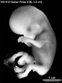
File:HillH13 Fetus bf01.jpg ==Human Fetus (Week 9)== HillH13 fetus week 9 CRL 3.8 cm left view.(1,200 × 1,600 (171 KB)) - 08:41, 14 December 2014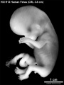
File:HillH13 Fetus bf02.jpg ==Human Fetus (Week 9)== HillH13 fetus week 9 CRL 3.8 cm left view.(1,200 × 1,600 (170 KB)) - 08:40, 14 December 2014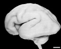
File:Human Fetus CRL240mm brain.jpg ==Human Fetus Brain== [[Category:Human Fetus]][[Category:Neural]](1,280 × 1,030 (129 KB)) - 16:05, 27 March 2018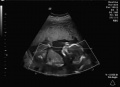
File:Ultrasound Fetus 01-icon.jpg (659 × 480 (39 KB)) - 16:40, 12 March 2013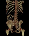
File:Fetus 35 week CT.jpg [[Category:Human Fetus]] [[Category:Computed Tomography]](700 × 874 (62 KB)) - 14:21, 24 August 2010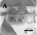
File:Dog fetus day 28.jpg ==Dog Fetus day 28==(400 × 375 (24 KB)) - 16:38, 25 September 2012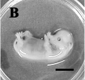
File:Dog fetus day 30.jpg ==Dog Fetus day 28==(400 × 375 (32 KB)) - 16:39, 25 September 2012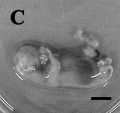
File:Dog fetus day 32.jpg ==Dog Fetus day 28==(400 × 375 (28 KB)) - 16:40, 25 September 2012
File:Human week 10 fetus 05.jpg ==Human Female Fetus - Heart (10 week)== {{Human Female Fetus Week 10 gallery}}(1,600 × 1,200 (612 KB)) - 16:39, 25 May 2016
File:Human week 10 fetus 06.jpg ==Human Female Fetus - Midgut Herniation (10 week)== {{Human Female Fetus Week 10 gallery}}(1,200 × 900 (251 KB)) - 17:01, 25 May 2016
File:Human week 10 fetus 23.jpg ==Human Female Fetus - Pelvic Region (10 week)== {{Human Female Fetus Week 10 gallery}}(1,600 × 1,200 (393 KB)) - 17:21, 25 May 2016
File:Human week 10 fetus 07.jpg ==Human Female Fetus - Spleen (10 week)== {{Human Female Fetus Week 10 gallery}}(1,200 × 900 (283 KB)) - 11:56, 30 May 2016
File:Human week 10 fetus 26.jpg ==Human Female Fetus - Midgut Herniation (10 week)== {{Human Female Fetus Week 10 gallery}}(1,200 × 900 (262 KB)) - 17:03, 25 May 2016
File:Human week 10 fetus 08.jpg ==Human Female Fetus Epiglottis (10 week)== {{Human Female Fetus Week 10 gallery}}(1,200 × 900 (323 KB)) - 21:03, 8 October 2015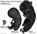
File:Size comparison embryo-fetus actual.jpg ==Size Comparison of the Embryo to Fetus== Original file name: EFsizeactual.jpg (Size comparison embryo-fetus actual.jpg)(194 × 178 (14 KB)) - 17:45, 6 May 2011
File:Human week 10 fetus 09.jpg ==Human Female Fetus - Atlas and Axis (10 week)== {{Human Female Fetus Week 10 gallery}}(1,200 × 900 (345 KB)) - 14:54, 25 May 2016
File:Human week 10 fetus 10.jpg ==Human Female Fetus - Pituitary and Lamina Terminalis (10 week)== {{Human Female Fetus Week 10 gallery}}(1,200 × 900 (291 KB)) - 09:21, 20 February 2019
File:Human week 10 fetus 11.jpg ==Human Female Fetus - Sacrum (10 week)== {{Human Female Fetus Week 10 gallery}}(1,200 × 900 (304 KB)) - 15:24, 25 May 2016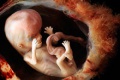
File:Fetus week 9-10-icon.jpg (220 × 147 (12 KB)) - 12:57, 24 April 2014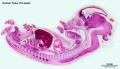
File:Human week 10 fetus 01.jpg ==Human Female Fetus (10 week)== {{Human Female Fetus Week 10 gallery}}(2,300 × 1,327 (448 KB)) - 11:57, 30 May 2016
File:Human week 10 fetus 12.jpg ==Human Female Fetus - Olfactory Nerve (10 week)== {{Human Female Fetus Week 10 gallery}}(1,200 × 900 (349 KB)) - 14:38, 25 May 2016
File:Human week 10 fetus 02.jpg ==Human Female Fetus (10 week)== See also [[:File:Human week 10 fetus 01.jpg|'''Large Image Version''']](800 × 462 (83 KB)) - 14:38, 11 October 2015
File:Human week 10 fetus 03.jpg ==Human Female Fetus - Pelvic Region (10 week)== {{Human Female Fetus Week 10 gallery}}(1,600 × 1,200 (370 KB)) - 17:21, 25 May 2016
File:Human week 10 fetus 04.jpg ==Human Female Fetus - Oral Cavity (10 week)== {{Human Female Fetus Week 10 gallery}}(1,600 × 1,200 (534 KB)) - 15:48, 25 May 2016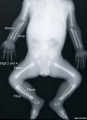
File:Human fetus skeleton x-ray 01.jpg ==Human Fetus Skeleton X-ray== {{Human fetus skeleton x-ray links}}(662 × 910 (54 KB)) - 17:05, 22 April 2014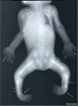
File:Human fetus skeleton x-ray 02.jpg ==Human Fetus Skeleton X-ray== Fetus with limb deformities including bowing of the digits, bowing and hypoplasia(686 × 928 (70 KB)) - 17:05, 22 April 2014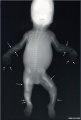
File:Human fetus skeleton x-ray 03.jpg ==Human Fetus Skeleton X-ray== Fetus with limb deformities (indicated by arrows), which include bowing of the ra(628 × 934 (81 KB)) - 17:04, 22 April 2014
File:2015BGDA Lecture - Development of the Embryo-Fetus 1.pdf ==BGDA Lecture - Development of the Embryo-Fetus 1== [[BGDA Lecture - Development of the Embryo/Fetus 1|Lecture Page]](2.65 MB) - 11:15, 7 May 2015File:2017 BGDA Lecture - Development of the Embryo-Fetus 1.pdf (3.39 MB) - 16:38, 3 May 2017File:2018 BGDA Lecture - Development of the Embryo-Fetus 1.pdf (8.38 MB) - 08:08, 4 May 2018File:2017 BGDA Lecture - Development of the Embryo-Fetus 2.pdf PDF version of 2017 lecture page: [[BGDA Lecture - Development of the Embryo/Fetus 2]](3.23 MB) - 08:44, 15 May 2017File:2018 BGDA Lecture - Development of the Embryo-Fetus 2.pdf [[BGDA Lecture - Development of the Embryo/Fetus 2]](5.67 MB) - 21:58, 13 May 2018File:2019 BGDA Lecture - Development of the Embryo-Fetus 1.pdf ==BGDA Lecture - Development of the Embryo/Fetus 1== [[BGDA Lecture - Development of the Embryo/Fetus 1]](1.93 MB) - 22:19, 2 May 2019File:BGDA Lecture 2014 - Development of the Embryo-Fetus 1.pdf ==BGDA Lecture - Development of the Embryo/Fetus 1==(869 KB) - 14:24, 7 May 2014File:2019 BGDA Lecture - Development of the Embryo-Fetus 2.pdf (3.59 MB) - 09:35, 13 May 2019


Page text matches

File:Koala fetus.jpg ==Koala Fetus== Koala fetus near birth.(559 × 1,000 (77 KB)) - 16:32, 1 December 2010File:HillH13 Fetus bf01.jpg ==Human Fetus (Week 9)== HillH13 fetus week 9 CRL 3.8 cm left view.(1,200 × 1,600 (171 KB)) - 08:41, 14 December 2014File:HillH13 Fetus bf02.jpg ==Human Fetus (Week 9)== HillH13 fetus week 9 CRL 3.8 cm left view.(1,200 × 1,600 (170 KB)) - 08:40, 14 December 2014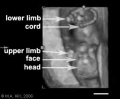
File:Ultrasound12wk 3D image.jpg ...ultrasound static image of the 12 week fetus shows a ventral view with the fetus upside down, with the head down and cord to the top. ...ategory:Genetic Abnormalities]] [[Category:Human Embryo]] [[Category:Human Fetus]](301 × 248 (8 KB)) - 15:10, 11 October 2009
File:HillH13 Fetus.gif ==Human Fetus (Week 9)== HillH13 fetus week 9 CRL 3.8 cm left view. (stereo pair animated gif)(450 × 600 (274 KB)) - 08:40, 14 December 2014File:Size comparison embryo-fetus actual.jpg ==Size Comparison of the Embryo to Fetus== Original file name: EFsizeactual.jpg (Size comparison embryo-fetus actual.jpg)(194 × 178 (14 KB)) - 17:45, 6 May 2011
File:Lineback1920 fig06-7.jpg ===Fig. 6. Cross-section of the ascending colon of a human fetus=== Cross-section of the ascending colon of a human fetus 105 mm. CR. length, showing the three tseniaj in a triangular position in t(1,200 × 808 (177 KB)) - 09:13, 17 January 2013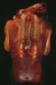
File:Spina Bifida 1.jpg ==Spina Bifida Fetus== Dorsal view of fetus with extensive spina bifida and anencephaly.(393 × 599 (49 KB)) - 10:28, 11 May 2016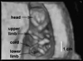
File:Ultrasound12wk 3D image2.jpg ==Ultrasound Fetus (12 week)== Image from near end of movie showing ventral view of fetus head to top, upper limbs, lower limbs and umbilical cord visible.(362 × 264 (8 KB)) - 09:06, 6 November 2012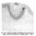
File:Keibel Mall 211.jpg ==Fig. 211 Human Fetus Integumentary Gland==(427 × 436 (43 KB)) - 13:01, 24 August 2012File:Human week 10 fetus 02.jpg ==Human Female Fetus (10 week)== See also [[:File:Human week 10 fetus 01.jpg|'''Large Image Version''']](800 × 462 (83 KB)) - 14:38, 11 October 2015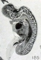
File:Mall Meyer1921 fig185.jpg ==Fig. 185. Fetus in sagittal section showing maceration== Fetus in sagittal section showing maceration, especially of the nervous system. N(341 × 500 (36 KB)) - 09:23, 13 December 2012
File:Fetal corpus cavernosum and corpus spongiosum 01.jpg * A - Corpus cavernosum, fetus of 13 WPC * C - Corpus cavernosum, fetus of 22 WPC(1,795 × 2,082 (919 KB)) - 11:42, 7 September 2014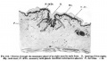
File:Keibel Mall 212.jpg ==Fig. 212 Human Fetus Integumentary Gland== Fetus 8 months.(650 × 371 (39 KB)) - 13:00, 24 August 2012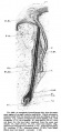
File:Keibel Mall 209.jpg ==Fig. 209 Human Fetus Integumentary Hair== Fetus Month 8.(458 × 990 (89 KB)) - 13:02, 24 August 2012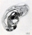
File:Mall Meyer1921 fig180.jpg ==Fig. 180. Fetus showing continuity of epidermis across the mouth== Fetus showing continuity of epidermis across the mouth, with obliteration of the(442 × 478 (32 KB)) - 09:20, 13 December 2012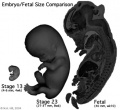
File:Size comparison embryo-fetus.jpg ==Size comparison of the Embryo to Fetus (week 10)== Original file name: EFsize.jpg (Size comparison embryo-fetus.jpg)(327 × 300 (29 KB)) - 17:46, 6 May 2011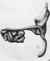
File:Bardeen1914-fig04.jpg ==Fig. 4. Gastrointestinal Tract Fetus 35 mm lateral view == ...ch, small and large intestines, rectum, bursa omentalis and mesentery of a fetus 35 mm long (No. 8, Wisconsin Collection).(840 × 1,019 (93 KB)) - 08:30, 29 April 2017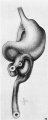
File:Bardeen1914-fig03.jpg ==Fig. 3. Gastrointestinal Tract Fetus 35 mm ventral view == ...ch, small and large intestines, rectum, bursa omentalis and mesentery of a fetus 35 mm long (No. 8, Wisconsin Collection).(405 × 1,014 (55 KB)) - 08:31, 29 April 2017File:Human week 10 fetus 07.jpg ==Human Female Fetus - Spleen (10 week)== {{Human Female Fetus Week 10 gallery}}(1,200 × 900 (283 KB)) - 11:56, 30 May 2016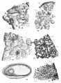
File:Simkins1928 plate09.jpg 38 Peripheral portion of an ovary from a seven-month fetus. X 750. 39 Peripheral portion of an ovary from a full-term fetus. cl.g., genitaloid cell. X 7 50.(1,548 × 2,096 (263 KB)) - 16:58, 31 January 2018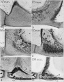
File:Anson1948 fig01.jpg (a) Base at posterior crus, left ear; 167 mm. fetus (Wisconsin series 105, slide 19, section 9). (b) Base and anterior crus, left ear; 175 mm. fetus (Wisconsin series 104, slide 17, section 5).(1,280 × 1,635 (310 KB)) - 17:02, 13 October 2017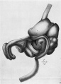
File:Bardeen1914-fig06.jpg ==Fig. 6. Lateral view fetus 40 mm== ...he stomach, small and large intestines, bursa omentalis and mesentery of a fetus 40 mm. long (No. 8, Wisconsin Collection).(721 × 1,000 (88 KB)) - 09:52, 3 October 2017File:2019 BGDA Lecture - Development of the Embryo-Fetus 1.pdf ==BGDA Lecture - Development of the Embryo/Fetus 1== [[BGDA Lecture - Development of the Embryo/Fetus 1]](1.93 MB) - 22:19, 2 May 2019File:2015BGDA Lecture - Development of the Embryo-Fetus 1.pdf ==BGDA Lecture - Development of the Embryo-Fetus 1== [[BGDA Lecture - Development of the Embryo/Fetus 1|Lecture Page]](2.65 MB) - 11:15, 7 May 2015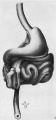
File:Bardeen1914-fig05.jpg ==Fig. 5. Ventral view fetus 40 mm== ...he stomach, small and large intestines, bursa omentalis and mesentery of a fetus 40 mm. long (No. 8, Wisconsin Collection). In [[:File:Bardeen1914-fig06.jpg(465 × 1,000 (61 KB)) - 07:05, 15 November 2015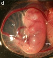
File:Wallaby embryo 05.jpg ==Tammar Wallaby Embryo and Fetus== ...ld within the folds of the yolk sac, which has become highly vascular. The fetus has well developed fore limbs ready for the climb to the pouch and the tong(996 × 1,100 (127 KB)) - 11:06, 29 July 2019
File:Mall Meyer1921 fig133.jpg ==Fig. 133. Marked macerated and deformed fetus from the same case== Fig. 133. Marked macerated and deformed fetus from the same case. X4.(365 × 610 (40 KB)) - 09:12, 6 December 2012
File:Mall Meyer1921 plate19.jpg Fig. 218. A fetus and cord showing marked maceration changes. No. 797. X1.35. Fig. 219. An older fetus, showing bleb-formation and curvature in extremities, due to maceration and(939 × 1,200 (237 KB)) - 18:44, 23 November 2012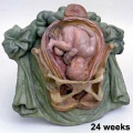
File:Galletti1770 week 24.jpg Fetus 24 weeks(450 × 450 (40 KB)) - 15:12, 13 October 2009
File:B050966-01.jpg ==Female Human Fetus 14cm==(2,000 × 992 (681 KB)) - 11:22, 25 June 2015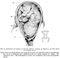
File:Frazer006 bw600.jpg ==Fetus during Third Trimester in Uterus compared to Non-pregnant Uterus== [[Category:Historic Embryology]] [[Category:Human Fetus]] [[Category:Third Trimester]] [[Category:Cartoon]](600 × 575 (47 KB)) - 17:30, 1 June 2013File:Human fetus skeleton x-ray 02.jpg ==Human Fetus Skeleton X-ray== Fetus with limb deformities including bowing of the digits, bowing and hypoplasia(686 × 928 (70 KB)) - 17:05, 22 April 2014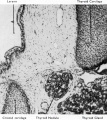
File:Boyd1950 fig09.jpg ==Fig. 9. Section fetus 145 mm to show aberrant portion of thyroid tissue lying between thyroid and {{Online Editor}} - [[Fetal Development|Fetus]] 145mm is about 16 weeks ({{GA}} 18 weeks).(801 × 900 (163 KB)) - 08:29, 21 March 2017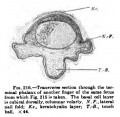
File:Keibel Mall 216.jpg ==Fig. 216 Human Fetus Finger== Transverse section through the terminal phalanx of another finger of the same fetus from which [[:File:Keibel Mall 215.jpg|Fig. 215]] is taken. The basal cell(366 × 356 (30 KB)) - 12:43, 13 January 2019File:Human week 10 fetus 01.jpg ==Human Female Fetus (10 week)== {{Human Female Fetus Week 10 gallery}}(2,300 × 1,327 (448 KB)) - 11:57, 30 May 2016File:Human fetus skeleton x-ray 03.jpg ==Human Fetus Skeleton X-ray== Fetus with limb deformities (indicated by arrows), which include bowing of the ra(628 × 934 (81 KB)) - 17:04, 22 April 2014
File:Mall Meyer1921 plate14.jpg Fig. 145. Enlarged fetus from same, showing maceration effects. X2. Fig. 150. Macerated, distorted normal cat fetus 10 mm. long. (After Kunz.)(941 × 1,200 (260 KB)) - 19:21, 23 November 2012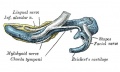
File:Gray0179.jpg ...outer aspect 95mm fetus]] | [[:File:Gray0181.jpg|Image - inner aspect 95mm fetus]](617 × 368 (47 KB)) - 18:34, 27 August 2012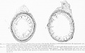
File:Lineback1920 fig04-5.jpg ==Cross-section of the colon of a human fetus== Fig. 4. Cross-section of the colon of a human fetus 52 mm. CR. length.(1,200 × 756 (150 KB)) - 09:16, 17 January 2013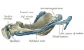
File:Gray0181.jpg ...outer aspect 95mm fetus]] | [[:File:Gray0181.jpg|Image - inner aspect 95mm fetus]] | [[Fetal Development]](617 × 368 (52 KB)) - 18:35, 27 August 2012File:Human Fetus CRL240mm brain.jpg ==Human Fetus Brain== [[Category:Human Fetus]][[Category:Neural]](1,280 × 1,030 (129 KB)) - 16:05, 27 March 2018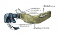
File:Gray0178.jpg ...outer aspect 95mm fetus]] | [[:File:Gray0181.jpg|Image - inner aspect 95mm fetus]](617 × 368 (44 KB)) - 18:33, 27 August 2012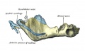
File:Gray0180.jpg ...outer aspect 95mm fetus]] | [[:File:Gray0181.jpg|Image - inner aspect 95mm fetus]] | [[Fetal Development]](617 × 368 (48 KB)) - 18:34, 27 August 2012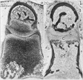
File:Anson1948 fig02.jpg ==Fig. 2. Photomicrographs of 202 mm and 310 mm fetus stapes and incus== ...0 (a) 202 mm fetus (Wisconsin series 70 slide 37, section 6); (b) 310 mm fetus (Wisconsin series 51, slide 38, section 6).(1,280 × 1,232 (241 KB)) - 08:55, 15 October 2017
File:Anson1948 fig07.jpg ==Fig. 7. Otic capsule developmental stages 210, 222, 275, 290 and 345 mm Fetus== ...fetus (Vlfisconsin series 59, slide 36, section 5); (e) 345 mm. (38 week) fetus (Wisconsin series 61,. slide 42, section 1).(1,280 × 1,801 (219 KB)) - 18:48, 13 May 2018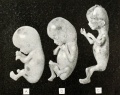
File:Streeter01.jpg '''A''', grade 1, fetus No. 1183; CR length 60 mm., weight 19.5 grams '''B''', grade 2, fetus No. 1282b, CR length 65.5 mm., weight I5 grams(800 × 634 (120 KB)) - 22:00, 12 May 2016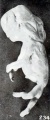
File:Mall Meyer1921 fig234.jpg ==Fig. 234. A very softened, macerated fetus==(331 × 793 (46 KB)) - 11:54, 3 December 2012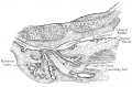
File:Lowsley1912 fig05.jpg ==Fig. 5. Human Fetus 16 cm Prostate==(1,641 × 1,083 (425 KB)) - 21:56, 16 June 2016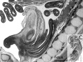
File:Human- fetal week 10 urogenital C.jpg ==Human Fetus (week 10) Female== Female fetus, 10 week, 40 mm CRL, early fetal, sagittal section, pelvic region(600 × 450 (105 KB)) - 17:42, 28 May 2011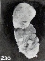
File:Mall Meyer1921 fig230.jpg ==Fig. 230. A decidedly mummified fetus==(281 × 379 (21 KB)) - 11:50, 3 December 2012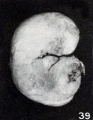
File:Mall Meyer1921 fig39.jpg ==Fig. 39. Macerated firmly rolled-up young fetus== The specimen consists of a rather poorly preserved young fetus, measuring 16.5 mm. CR. The ventral abdominal wall had been seriously injur(414 × 535 (41 KB)) - 07:11, 4 December 2012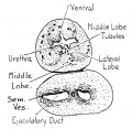
File:Lowsley1912 fig02.jpg ==Fig. 2. Human Fetus 7.5 cm==(759 × 740 (102 KB)) - 21:52, 16 June 2016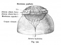
File:Kollmann722.jpg pupillaris sind nach einem menschlichen Fetus von 8 Monaten, die Membrana capsularis nach einem menschlichen Fetus von 6 Wochen eingezeichnet; +:(533 × 391 (28 KB)) - 10:52, 21 October 2011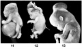
File:Mall1917 fig11-13.jpg '''Fig. 11''' Stunted fetus with a large hernia in umbilical cord, also spina bifida. '''Fig. 13''' Normal fetus with hernia of midbrain.(1,000 × 599 (79 KB)) - 06:04, 12 November 2013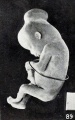
File:Mall Meyer1921 fig89.jpg ==Fig. 89. Normal fetus with hernia of mid-brain==(535 × 854 (90 KB)) - 08:24, 3 December 2012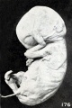
File:Mall Meyer1921 fig176.jpg ==Fig. 176. Fetus showing gluing of hand to face==(537 × 807 (70 KB)) - 09:18, 13 December 2012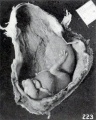
File:Mall Meyer1921 fig223.jpg ==Fig. 223. External appearance of fetus in situ==(562 × 705 (80 KB)) - 11:43, 3 December 2012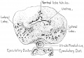
File:Lowsley1912 fig06.jpg ==Fig. 6. Human Fetus 19 cm Prostate==(1,327 × 916 (237 KB)) - 21:57, 16 June 2016
File:McMurrich1930 fig85.jpg ==Fig. 85. Representations of human fetus at term and of ungulate placenta== .... Where the uterus is also shown there is usually no indication of how the fetus is connected with it; only in QIII, 8 are the cotyledons represented, on th(1,280 × 1,698 (373 KB)) - 11:46, 20 April 2020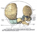
File:Keibel Mall 321.jpg ==Fig. 321 Lateral view of the cranium of a human fetus 80 mm long== Lateral view of the cranium of a human fetus 80 mm long.(746 × 651 (122 KB)) - 19:33, 4 September 2014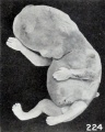
File:Mall Meyer1921 fig224.jpg ==Fig. 224. An older macerated fetus with extremities extended instead of folded==(440 × 551 (43 KB)) - 11:44, 3 December 2012
File:Mall Meyer1921 fig210.jpg ==Fig. 210. Normal well-preserved cat fetus==(240 × 379 (17 KB)) - 10:14, 3 December 2012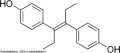
File:Diethylstilbestrol.jpg * Female fetus - increased risk abnormal reproductive tract and cancer. * Male fetus - abnormal genitalia.(600 × 263 (16 KB)) - 11:29, 6 March 2019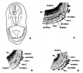
File:Humphrey1940 fig04.jpg ==Fig. 4. Olfactory and accessory olfactory formations in the 37 mm fetus== ...illustrate the olfactory and accessory olfactory formations in the 37 mm. fetus. Activated protargol preparation.(1,000 × 882 (101 KB)) - 17:50, 24 October 2017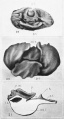
File:Atwell1926 plate06.jpg 19 Wax—plalte reconstruction of the hypophysis from :1. 45-mm. human fetus (Huber collection, no. XVIII), viewed from above and in front. X 35. 20 Reconstruction of the epithelial hypophysis from a 55»mm. human fetus (U.B.E.C., no. 25), viewed from above and in front. X 35.(809 × 1,500 (136 KB)) - 15:09, 9 November 2016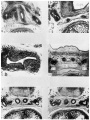
File:Watson1918 plate01.jpg ...ion, 110. 1358e). A graphic reconstruction of these structures in the same fetus is shown in text figure 4. In figure 10 the left deferent duct is cut obl ..., estimated age 19 weeks (Carnegie Collection, no. 1049). This is the same fetus shown in text-figure 6. In figure 15 it will be noted that the left ampul(1,200 × 1,601 (398 KB)) - 09:21, 8 December 2016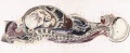
File:BrauneB2.jpg ==The Position of the Uterus and Fetus at Term (1872)== ...tial section through the fetus shows the relative size and position of the fetus during pregnancy.(1,200 × 485 (143 KB)) - 22:57, 3 June 2015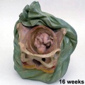
File:Galletti1770 week 16.jpg ==Fetus 16 weeks==(450 × 450 (32 KB)) - 08:00, 27 May 2010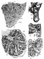
File:Simkins1928 plate03.jpg 14 Cross~section of the testis of a fetus, 190 mm. X 40. 15 Detail of the tubules of a 190-mm. fetus. X 750.(1,551 × 2,086 (293 KB)) - 16:45, 31 January 2018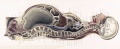
File:BrauneA1.jpg ==The Position of the Uterus and Fetus at Term (1872)== ...tial section through the fetus shows the relative size and position of the fetus during pregnancy.(1,200 × 485 (141 KB)) - 17:13, 30 October 2012File:Human week 10 fetus 05.jpg ==Human Female Fetus - Heart (10 week)== {{Human Female Fetus Week 10 gallery}}(1,600 × 1,200 (612 KB)) - 16:39, 25 May 2016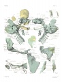
File:Macklin-plate2a.jpg ==Plate 2. The skull of a human fetus of 43 millimeters greatest length==(1,142 × 1,500 (361 KB)) - 16:45, 23 April 2014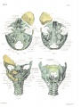
File:Macklin-plate1a.jpg ==Plate 1. The skull of a human fetus of 43 millimeters greatest length==(1,142 × 1,500 (374 KB)) - 16:45, 23 April 2014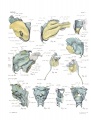
File:Macklin-plate5a.jpg ==Plate 5. The skull of a human fetus of 43 millimeters greatest length==(1,142 × 1,500 (324 KB)) - 16:46, 23 April 2014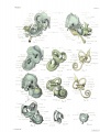
File:Macklin-plate3a.jpg ==Plate 3. The skull of a human fetus of 43 millimeters greatest length==(1,142 × 1,500 (321 KB)) - 16:46, 23 April 2014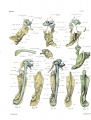
File:Macklin-plate4a.jpg ==Plate 4. The skull of a human fetus of 43 millimeters greatest length==(1,142 × 1,500 (335 KB)) - 16:46, 23 April 2014
File:Lowsley1912 fig01.jpg ==Fig 1. Human Fetus 5 cm==(990 × 620 (29 KB)) - 21:53, 16 June 2016File:Human week 10 fetus 12.jpg ==Human Female Fetus - Olfactory Nerve (10 week)== {{Human Female Fetus Week 10 gallery}}(1,200 × 900 (349 KB)) - 14:38, 25 May 2016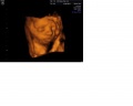
File:Ultrasound Demonstrating Facial Features.jpg ...ere the face of the fetus is clearly shown. This is an example of a normal fetus.(1,280 × 1,024 (61 KB)) - 19:50, 5 October 2011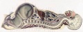
File:BrauneC2.jpg ==The Position of the Uterus and Fetus at Term (1872)== ...n through the maternal anatomy shows the relative size and position of the fetus during pregnancy.(1,200 × 461 (142 KB)) - 17:13, 30 October 2012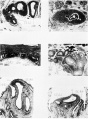
File:Watson1918 plate02.jpg ...th, estimated age 21 weeks (Carnegie Collection, no. 1171), being the same fetus shown in text figure 7. In figure 16 is shown the left anipulla and seminal '''18 to 21''' Transverse sections through the seminal tract in a human fetus 221 mm. crown-rump length, estimated age 25 weeks (Carnegie Collection, no.(1,200 × 1,628 (367 KB)) - 09:33, 8 December 2016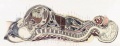
File:BrauneC3.jpg ==The Position of the Uterus and Fetus at Term (1872)== ...n through the maternal anatomy shows the relative size and position of the fetus during pregnancy.(1,200 × 461 (143 KB)) - 22:58, 3 June 2015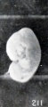
File:Mall Meyer1921 fig211.jpg ==Fig. 211. Normal poorly preserved cat fetus of approximately the same length==(212 × 470 (18 KB)) - 10:15, 3 December 2012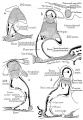
File:Anson1948 fig06.jpg ==Fig. 6. Otic capsule developmental stages 190 mm and 215 mm fetus== ...) fetus (Wisconsin series 129, slide 20, section 3); (d) 215 mm. (24 week) fetus (Wisconsin series 62, slide 28, section 4). Parts (1, b and d represent sec(1,028 × 1,488 (224 KB)) - 20:16, 16 October 2017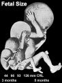
File:Fetal size change.jpg [[Category:Human Fetus]](337 × 443 (18 KB)) - 11:04, 16 August 2014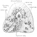
File:Lowsley1912 fig07.jpg ==Fig. 7. Human Fetus 27 cm Prostate==(1,437 × 1,435 (486 KB)) - 21:58, 16 June 2016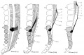
File:Streeter1919-fig02.jpg ...Topographical relations of the caudal end of the spinal cord in the human fetus from the eighth to the twenty-fifth week== ...long; 67-mm. fetus, 4.75 mm. long; 111-mm. fetus, 12.25 mm. long; 221-mm. fetus, 32 mm. long.(2,023 × 1,318 (499 KB)) - 22:24, 31 January 2019File:2018 BGDA Lecture - Development of the Embryo-Fetus 2.pdf [[BGDA Lecture - Development of the Embryo/Fetus 2]](5.67 MB) - 21:58, 13 May 2018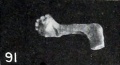
File:Mall Meyer1921 fig91.jpg From No. 230, a fetus compressus 57 mm. CR. X0.75.(352 × 191 (14 KB)) - 15:07, 15 January 2013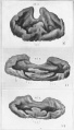
File:Atwell1926 plate05.jpg 16 Wax~platc reconstruction of the epithelial hypophysis from a. 25-mm. human fetus (U.B.E.C., no. 33), viewed from above and in front. X 65. 18 Reconstruction of the epithelial hypophysis from a 26-mm. human fetus (U.B.E.C., 110. 1), viewed from above and in front. X 50.(855 × 1,500 (135 KB)) - 16:30, 9 November 2016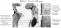
File:HansonAnson1962 fig04.jpg ==Fig. 4. Fetus 15 week 115 mm== '''a''', In the 15-week fetus (115 mm.) the malleus begins to ossify from a single center located medial(1,280 × 598 (201 KB)) - 10:31, 7 January 2019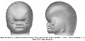
File:Keibel Mall 070-071.jpg ==Fig. 68-69 Head of a Fetus 47.5 mm==(1,000 × 490 (45 KB)) - 21:14, 10 September 2012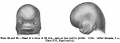
File:Keibel Mall 068-069.jpg ==Fig. 68-69 Head of a Fetus 25mm==(1,000 × 358 (35 KB)) - 21:14, 10 September 2012
File:Grays Anatomy Embryology cover.jpg Cover image for iBook developed from [[:File:Gray0038.jpg|Figure 38]] - Fetus in Utero between Fifth and Sixth Months.(459 × 600 (25 KB)) - 18:45, 13 May 2013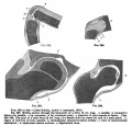
File:Keibel Mall 285-288.jpg Fig. 285. Median section through the knee-joint of a fetus 13 cm long. a, patella; b. connective tissue over patella; c, lig Fig. 286-288. Hip-joint of a male fetus 25 cm. long, of a female child six years old, and of a male adult. a. regio(703 × 674 (72 KB)) - 02:38, 14 September 2012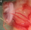
File:Wallaby embryo 04.jpg ==Tammar Wallaby Embryo and Fetus== Fetus at day 23 of pregnancy, showing the increase in the vascular region and the(1,138 × 1,098 (110 KB)) - 13:17, 23 May 2012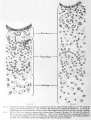
File:Streeter002-3.jpg ==Fig. 2. Detail of the lateral semicircular duct in a human fetus 30 mm. long== ==Fig. 3. Detail of the lateral canal in a human fetus 16 mm. long==(607 × 800 (106 KB)) - 12:41, 15 February 2011File:Human week 10 fetus 08.jpg ==Human Female Fetus Epiglottis (10 week)== {{Human Female Fetus Week 10 gallery}}(1,200 × 900 (323 KB)) - 21:03, 8 October 2015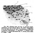
File:Keibel Mall 197.jpg ==Fig. 200 Human Fetus==(500 × 469 (46 KB)) - 07:15, 23 August 2012


