Search results
From Embryology
Page title matches
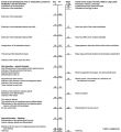
File:Octopus bimaculatus embryonic development timeline.jpg ==Embryonic Development of Octopus bimaculatus== ...e table compares the embryonic development with the scale proposed for the development of L. pealii (Arnold 1965). *Observed in vivo.(1,280 × 1,394 (187 KB)) - 11:02, 21 May 2020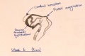
File:Week 6 embryonic development of CNS.jpg ==Week 6 embryonic development of CNS and emergence of pineal evagination.==(1,209 × 794 (165 KB)) - 12:43, 24 October 2014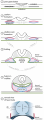
File:Development of Trilaminar Embryonic Disc .png (1) Trilaminalar embryonic disc after gastrulation; lateral plate mesoderm resides between the endoder (4) By folding the lateral edges of the embryonic disc move inwards, creating the anterior intestinal portal. The persisting(1,921 × 5,223 (1.24 MB)) - 10:44, 13 October 2017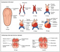
File:Progressive development of the Embryonic Heart.jpeg Development of the heart in the fetus and partitioning of the heart into four chambers ...gy, Connexions Web site. https://cnx.org/contents/FPtK1zmh@6.27:GxtDaPpg@3/Development-of-the-Heart/, Jun 19, 2013.(1,024 × 883 (154 KB)) - 11:30, 4 September 2018


Page text matches
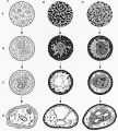
File:Hilfer1990 Fig16.jpg ==Figure 16. Townes and Holtfreter (1955) different embryonic layers in amphibians== ...o clumps and tended to take a position resembling that of normal embryonic development.(1,200 × 1,333 (247 KB)) - 10:43, 28 August 2014File:Octopus bimaculatus embryonic development timeline.jpg ==Embryonic Development of Octopus bimaculatus== ...e table compares the embryonic development with the scale proposed for the development of L. pealii (Arnold 1965). *Observed in vivo.(1,280 × 1,394 (187 KB)) - 11:02, 21 May 2020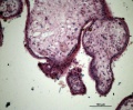
File:HillH52 chorionic villi 05.jpg Human early placental villi development at 4-5 weeks. (Hill H52, x20) {{HE}} scale bar - 100 μm) * Embryonic mesenchyme(1,200 × 992 (371 KB)) - 13:54, 22 March 2014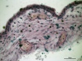
File:HillH52 chorionic villi 04.jpg Human early placental villi development at 4-5 weeks. (Hill H52, x40) {{HE}} scale bar - 50 μm) * Embryonic mesenchyme(1,200 × 900 (254 KB)) - 13:34, 22 March 2014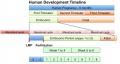
File:Human development timeline graph 02.jpg ==Human Development Timeline== |Simplified graphical view of human development timeline.(800 × 424 (61 KB)) - 09:37, 14 April 2016
File:Gray0032.gif ...mbryonic ectoderm that will contribute to embryonic and placental membrane development ...storic term for what we today call endoderm that will contribute to embryo development(500 × 417 (57 KB)) - 11:13, 22 May 2011
File:Bat embryo stage 19.mov ==Bat Embryonic Development (stage 19)== ...Cancer Center, who provided images and stage information on the embryonic development of the bat.(30 KB) - 01:12, 29 April 2011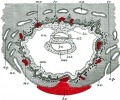
File:Gray0032.jpg ...ough showing the trilaminar embryo suggests late week 2 to early week 3 of development. Uterine cavity is shown at bottom of image. ...mbryonic ectoderm that will contribute to embryonic and placental membrane development(800 × 667 (159 KB)) - 08:51, 21 April 2013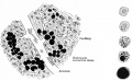
File:Bailey111.jpg ...the right are five individual cells showing stages of development from an embryonic cell to an adult fat cell.(916 × 567 (117 KB)) - 13:05, 18 January 2011
File:Stage13 and 22 thyroid development.jpg ==Thyroid Gland Embryonic Development Overview== Cross-sections of head and neck regions of embryonic stage 13 (week4-5) and stage 22 (week 8) are shown.(1,000 × 800 (283 KB)) - 11:49, 30 August 2011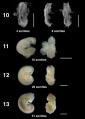
File:Bat embryo stage 10 to 13.jpg ==Bat Embryonic Development (stage 10-13)== ...Cancer Center, who provided images and stage information on the embryonic development of the bat.(800 × 1,122 (66 KB)) - 13:21, 6 July 2012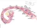
File:Mouse E0-E5.jpg ==Development of the Preimplantation Blastocyst in Mice== Cartoon shows an overview of mouse development from Embryonic Day 0 (E0) Through Day 5 (E5.0).(991 × 749 (90 KB)) - 10:08, 14 October 2016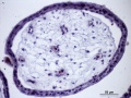
File:HillH52 chorionic villi 08.jpg Human early placental villi development at 4-5 weeks. (Hill H52, x40) {{HE}} scale bar - 50 μm) ...phoblast shell enclosing mesenchyme (extra-embryonic mesoderm), containing embryonic blood vessels.(1,200 × 900 (229 KB)) - 14:59, 3 July 2014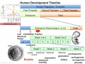
File:Human development timeline graph 01.jpg ==Human Development Timeline== This graph shows an overview of human development and details about embryonic development.(1,000 × 750 (141 KB)) - 08:49, 15 April 2014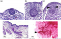
File:Mouse mammary development 01.jpg ==Embryonic Mouse Mammary Development== (a) [[:Category:Mouse E12.5|Embryonic day E12.5]] The epithelial cells have invaginated to form the initial bud,(1,200 × 773 (147 KB)) - 09:04, 20 March 2018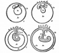
File:Kellicott 179.jpg From McMurrich (Development of the Human Body). ...ic ectoderm; the dotted line marks the line of the transition of the body (embryonic) ectoderm into that of the amnion.(893 × 800 (131 KB)) - 16:54, 23 December 2013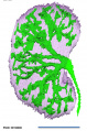
File:Human embryonic renal branching stage 22.jpg ==Embryonic Human Renal Urothelial Branching Development== ...he [[Kyoto Collection]] at Carnegie stage {{CS22}}. See also [[:File:Human embryonic renal branching 1.jpg|branching figure stages 14-22]].(500 × 756 (150 KB)) - 10:21, 18 January 2019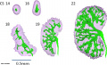
File:Human embryonic renal branching 1.jpg ==Embryonic Human Renal Urothelial Branching Development== ...{CS14}}, {{CS16}}, {{CS18}}, {{CS19}} and {{CS22}}. See also [[:File:Human embryonic renal branching stage 22.jpg|branching figure stage 22]].(1,280 × 779 (236 KB)) - 10:26, 18 January 2019- Error creating thumbnail: File with dimensions greater than 12.5 MP
File:Worm - embryonic cell lineage 01.jpg ==Worm - Embryonic Cell Lineage== Embryonic cell lineage developed by J .E. Sulston, E. Schierenberg, J. G. White, J. N(10,389 × 1,336 (598 KB)) - 11:25, 30 June 2012 
File:GladstoneHamilton1941 text-fig04.jpg ==Text-fig. 4. General .view of the embryonic disc at the anterior part of the primitive streak and groove== ...ion no. 57, the sections having been numbered from the anterior end of the embryonic disc. x 100.(1,280 × 633 (96 KB)) - 16:58, 26 February 2017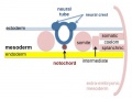
File:Mesoderm-cartoon4.jpg ==Mesoderm and Ectoderm Development cartoon== ...s a section through the trunk of the trilaminar embryo showing the further development of the 3 layers and the space (coelom) that forms in the mesoderm (only the(400 × 300 (20 KB)) - 10:09, 23 April 2020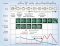
File:Model human blastocyst development.jpg ==Proposed Model for Human Embryo Development== ...gg (ESSP1) must be degraded as the transition from oocyte to embryo begins embryonic genome activation (EGA).(946 × 726 (84 KB)) - 14:47, 6 October 2015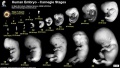
File:Human Carnegie stage 1-23.jpg ==Human Embryonic Development== ..., showing human external appearance and growth during the first 8 weeks of development.(1,000 × 563 (98 KB)) - 11:32, 14 May 2017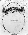
File:Stage6 bf03.jpg ...howing development of the bilaminar (two layer) embryo and the early extra-embryonic coeloms (spaces). Extra-embryonic coelom(646 × 800 (65 KB)) - 14:48, 1 October 2018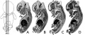
File:Human- fetal week 10 sagittal planes.jpg ...he embryonic period (up to week 8) but still only 2 weeks into early fetal development.(600 × 250 (29 KB)) - 15:39, 27 April 2010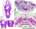
File:Stage13 and 22 thyroid development a.jpg ==Thyroid Gland Embryonic Development== ...e developing thyroid at the beginning of week 5 and at week 8 of embryonic development.(800 × 640 (94 KB)) - 08:50, 22 May 2012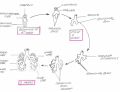
File:Lung development overiview.png Summary of the lung formation from the embryonic stage to the fetal stage. Original Image is adapted from: Figure 1 from Embryonic Development of the Respiratory System page(717 × 549 (178 KB)) - 14:59, 9 November 2014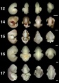
File:Bat embryo stage 12-17.jpg ==Bat Embryonic Development (stage 12-17)== :'''Links:''' [[Bat Development]] | [[:File:Bat embryo stage 10-13.jpg|stage 10 to 13]] | [[:File:Bat embry(548 × 767 (30 KB)) - 01:16, 29 April 2011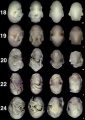
File:Bat embryo stage 18-24.jpg ==Bat Embryonic Development (stage 18-24)== :'''Links:''' [[Bat Development]] | [[:File:Bat embryo stage 10-13.jpg|stage 10 to 13]] | [[:File:Bat embry(518 × 734 (32 KB)) - 01:16, 29 April 2011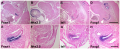
File:Thymic Epithelial Cell Development and Function.png ==Thymic Epithelial Cell Development and Function== '''Image shows Thymic Epithelial Cell Development and Function in fetal period'''(2,028 × 823 (3.17 MB)) - 11:37, 9 November 2014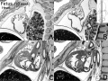
File:Human- fetal week 10 heart ABCD.jpg ...he embryonic period (up to week 8) but still only 2 weeks into early fetal development.(600 × 450 (133 KB)) - 16:10, 27 April 2010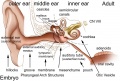
File:Adult hearing embryonic origins.jpg ==Adult Hearing Embryonic Origins== ...nd inner), each of which have their own separate components from different embryonic origins.(1,000 × 675 (80 KB)) - 14:26, 8 May 2018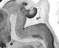
File:Human- fetal week 10 cerebellum C.jpg ...he embryonic period (up to week 8) but still only 2 weeks into early fetal development.(347 × 284 (25 KB)) - 18:53, 20 September 2012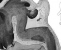
File:Human- fetal week 10 cerebellum D.jpg ...he embryonic period (up to week 8) but still only 2 weeks into early fetal development.(347 × 284 (23 KB)) - 18:53, 20 September 2012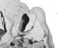
File:Human- fetal week 10 cerebellum B.jpg ...he embryonic period (up to week 8) but still only 2 weeks into early fetal development.(347 × 284 (21 KB)) - 18:53, 20 September 2012
File:BGDA PracManual 2011 Practical 7.pdf ==BGDA Practical Manual 2011 Practical 7 - Embryonic Development==(203 KB) - 14:43, 22 April 2011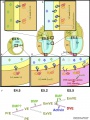
File:Mouse distal visceral endoderm 01.jpg ...MP signaling promotes the differentiation of primitive endoderm (PrE) into embryonic visceral endoderm (VE) until E4.5. (B) Embryonic visceral endoderm (VE) differentiates as a result of the concerted action o(959 × 1,280 (208 KB)) - 13:16, 3 May 2013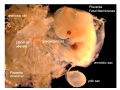
File:Placental membranes.jpg * '''amniotic sac''' - formed by the amniotic membrane (ectoderm and extra-embryonic mesoderm) completely surrounding the surrounding the embryo. * '''yolk sac''' - the yolk membrane (endoderm and extra-embryonic mesoderm) attached to the embryo at the umbilicus and continuous with the m(600 × 450 (99 KB)) - 07:46, 15 May 2014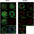
File:Antibody immunostaining of ligands and receptors in triploid human embryos.png ...mbryonic nuclei. Furthermore, growth factors were used to aid in embryonic development and promote blastocyst growth as well as IgG being applied to the negative ..., Shu Y, Cheng Y, Qiao J, et al. (2012) Promotion of Human Early Embryonic Development and Blastocyst Outgrowth In Vitro Using Autocrine/Paracrine Growth Factors.(3,439 × 3,444 (7.13 MB)) - 00:21, 20 August 2014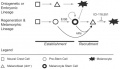
File:Zebrafish melanocyte development model.jpg ==Model for the parallel establishment of the zebrafish embryonic melanocyte lineage and the adult melanocyte stem cell lineage== ...and melanocytes during metamorphosis or regeneration, when inhibition from embryonic melanocytes (block arrow) is relieved. The MSC–derived melanocytes are se(484 × 277 (40 KB)) - 14:50, 20 February 2011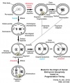
File:Aneuploidy model based on fragmentation.jpg ==Model for the Origin of Human Embryonic Aneuploidy Based on Fragmentation Timing== ...omal abnormalities and undergoes fragmentation as a survival mechanism. As development proceeds, these fragments either remain or are reabsorbed by the blastomere(668 × 790 (105 KB)) - 13:44, 1 April 2019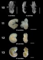
File:Bat embryo stage 10-13.jpg ==Bat Embryonic Development (stage 10-13)== :'''Links:''' [[Bat Development]] | [[:File:Bat embryo stage 10-13.jpg|stage 10 to 13]] | [[:File:Bat embry(547 × 767 (23 KB)) - 01:34, 29 April 2011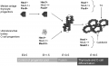
File:ThyroidDevelopment.png This image summarises the endodermal and mesodermal contribution to the development of the thyroid gland. The progenitor cells are from anterior endoderm and r ...ed to understand what has gone before, you have not included much in fetal development. There is no explanation of the molecular factors shown in the figure.(1,996 × 1,212 (49 KB)) - 11:46, 9 November 2014File:Human blastocyst day 3-6.mov ...asive imaging of human embryos before embryonic genome activation predicts development to the blastocyst stage. ...asive imaging of human embryos before embryonic genome activation predicts development to the blastocyst stage(4.26 MB) - 16:08, 15 October 2010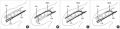
File:Embryonic upper limb - brachial and superficial brachial artery.jpg ==Embryonic upper limb - brachial and superficial brachial artery == ...development of the brachial artery and the superficial brachial artery in embryonic upper limb.(1,280 × 314 (122 KB)) - 14:25, 2 February 2020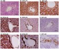
File:Histology of human embryonic liver at 11 weeks.png ==Histology of human embryonic liver at 11 weeks== Paraffin-embedded sections of human embryonic liver at 11 weeks (9 weeks of gestation) stained for Hematoxylin and Eosin(1,988 × 1,642 (4.17 MB)) - 22:02, 8 November 2014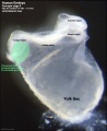
File:Stage9 bf2-primordial germ cell region.jpg # the relative size of the embryo and the associated extra-embryonic coeloms. # the shape of the early folded embryonic disc and rostro-caudal bendings.(814 × 1,000 (72 KB)) - 21:20, 17 May 2015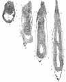
File:Bailey061.jpg ...ys and 22 hours after insemination (younger than B but further advanced in development), showing beginning of proamniotic cavity. ...astocyst 8 days after insemination (younger than B but further advanced in development), showing more advanced proamniotic cavity.(878 × 1,086 (184 KB)) - 15:02, 26 January 2011
File:1 Min Embryo - Human timeline.mp4 ...mplantation to 8 Weeks|BGDA Prac 6]] | [[Embryonic Development]] | [[Fetal Development]] | [[One Minute Embryology]] Lets look at an overview of human development(5.28 MB) - 10:40, 28 April 2016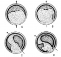
File:Rugh 077.jpg ...r V. Hamburger and B. Mayer, unpublished. Redrawn from Spemann: "Embryonic Development and Induction," New Haven, Yale University Press.(861 × 800 (171 KB)) - 13:43, 12 April 2013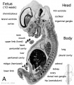
File:Human- fetal week 10 sagittal plane A.jpg ...he embryonic period (up to week 8) but still only 2 weeks into early fetal development.(500 × 573 (96 KB)) - 16:19, 27 April 2010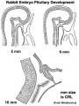
File:Pituitary rabbit development.jpg ==Rabbit Pituitary Development== Cartoon showing the changes in the embryonic rabbit pituitary.(374 × 500 (33 KB)) - 14:52, 27 May 2014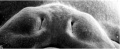
File:Stage19 em01.jpg ...EM images focussing on this developmentally important region and time for embryonic cleft lip and palate.(800 × 329 (37 KB)) - 08:17, 23 February 2014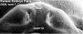
File:Stage19 em11.jpg ...EM images focussing on this developmentally important region and time for embryonic cleft lip and palate.(800 × 329 (46 KB)) - 10:11, 23 February 2014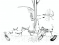
File:Grasshopper lifecycle.jpg # As soon as the eggs are laid, they begin embryonic development and reach an advanced stage in which they enter diapause and pass the winte # In spring the eggs complete embryonic development and hatch.(1,072 × 814 (122 KB)) - 13:40, 16 February 2016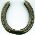
File:Horseshoe.jpg ...]] within the lateral plate [[mesoderm]] that forms during week 3 of human development. ...:''' [[Renal_System_-_Abnormalities|Renal Abnormalities]] | [[Renal System Development]](400 × 400 (32 KB)) - 09:09, 13 October 2016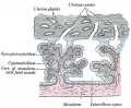
File:Gray0037.jpg ...evelopment contain core of mesoderm. Tertiary villi then have blood vessel development within this core. Extra-embryonic mesoderm grows into villi, covers the entire surface of chorionic sac.(500 × 412 (74 KB)) - 11:21, 9 June 2014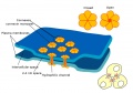
File:Gap junction 01.jpg * Also in embryonic development (see [[Blastocyst Development]])(800 × 562 (69 KB)) - 12:35, 25 March 2015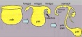
File:Endoderm cartoon.jpg ==Cartoon of endoderm development== ...3 images is from the animation [[Development_Animation_-_Endoderm|Endoderm Development]](587 × 262 (31 KB)) - 19:42, 11 June 2013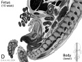
File:Human- fetal week 10 lower body D.jpg ...he embryonic period (up to week 8) but still only 2 weeks into early fetal development.(600 × 450 (91 KB)) - 15:36, 27 April 2010File:Stages 1-5 mouse.pdf Table 1: Mouse embryonic stages of development from fertilization to zona free blastocyst (stages 1 to 5).(29 KB) - 13:42, 31 March 2012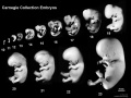
File:Human Carnegie stage 10-23.jpg ==Human Embryonic Development== ...tion, embryos compiled and scaled from original images. Note that weeks of development are form fertilisation (fertilization age), not gestational age {{GA}} that(1,024 × 768 (95 KB)) - 11:38, 16 February 2018
File:Elephant and calf.jpg * 50 days - embryonic vesicle earliest observation * 71 days - embryonic heartbeat and allantois visible as a single sacculation(538 × 404 (52 KB)) - 20:24, 1 November 2013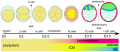
File:Morphological differences in early mouse embryonic development.png ...hological and lineage specification steps during the early mouse embryonic development== ...is formed as a layer separating epiblast from blastocoel (E4.5). After 4.5 embryonic days, the preimplantation embryo contains more than 100 cells.(2,061 × 886 (455 KB)) - 12:47, 18 August 2016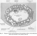
File:Hamilton1949 fig12.jpg ==Fig.12. Diagram to show the development of the secondary yolk sac, extra-embryonic coelom and remnant of the primary yolk sac==(1,312 × 1,265 (267 KB)) - 15:30, 15 May 2018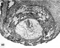
File:Hertig1956 fig82.jpg ...ssible to say what effect, if any, such an abnormality would have upon the development of the body stalk and the embryo. [[:Category:Carnegie Embryo 8299|Carnegie(1,000 × 796 (181 KB)) - 14:37, 25 February 2017
File:Human- fetal week 10 sagittal plane B.jpg ...he embryonic period (up to week 8) but still only 2 weeks into early fetal development.(500 × 573 (99 KB)) - 15:49, 27 April 2010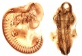
File:Bat - neural development 01.jpg ==Bat neural Development (stage 14)== :'''Links:''' [[Bat Development]] | [[Neural System Development]] | [[Carnegie stage 14]](733 × 498 (33 KB)) - 13:17, 6 July 2012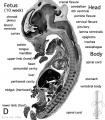
File:Human- fetal week 10 sagittal plane D.jpg ...he embryonic period (up to week 8) but still only 2 weeks into early fetal development.(500 × 573 (105 KB)) - 07:41, 1 May 2011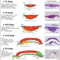
File:Cattle embryo staging 01.jpg ...al sections (H&E or CER1 stained) of, cattle embryos at the five stages of development between hatching and the start of {{gastrulation}}, based on data from sect ...ural) hypoblast; PS, primitive streak; RL, Rauber’s Layer (polar TB); VH, (embryonic) visceral hypoblast. All bars are 100 μm.(1,961 × 1,962 (708 KB)) - 13:45, 22 November 2018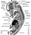
File:Human- fetal week 10 sagittal plane C.jpg ...he embryonic period (up to week 8) but still only 2 weeks into early fetal development.(500 × 573 (98 KB)) - 15:48, 27 April 2010
File:Hans Spemann.jpg A German embryologist who worked extensively on amphibian development and was the discoverer of the organiser region (or primitive node) the cont ...iology or Medicine "for his discovery of the organizer effect in embryonic development".(300 × 425 (18 KB)) - 10:48, 17 September 2014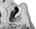
File:Human- fetal week 10 cerebellum A.jpg ...he embryonic period (up to week 8) but still only 2 weeks into early fetal development.(347 × 284 (24 KB)) - 18:54, 20 September 2012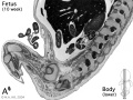
File:Human- fetal week 10 lower body A.jpg ...he embryonic period (up to week 8) but still only 2 weeks into early fetal development.(600 × 450 (96 KB)) - 15:52, 27 April 2010
File:Human- fetal week 10 urogenital A.jpg ...he embryonic period (up to week 8) but still only 2 weeks into early fetal development.(600 × 450 (109 KB)) - 15:52, 27 April 2010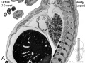
File:Human- fetal week 10 upper body A.jpg ...he embryonic period (up to week 8) but still only 2 weeks into early fetal development.(600 × 450 (104 KB)) - 15:53, 27 April 2010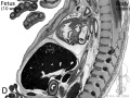
File:Human- fetal week 10 upper body D.jpg ...he embryonic period (up to week 8) but still only 2 weeks into early fetal development.(600 × 450 (106 KB)) - 15:42, 27 April 2010
File:Human embryonic-fetal tongue 01.jpg Photographs of mid-sagittal sections of an embryonic human tongue. (A, B) Tongue development (TD) stage 1. Co, copula; Ce, foramen cecum.(1,000 × 1,129 (490 KB)) - 19:02, 18 June 2018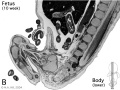
File:Human- fetal week 10 lower body B.jpg ...he embryonic period (up to week 8) but still only 2 weeks into early fetal development.(600 × 450 (93 KB)) - 15:51, 27 April 2010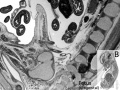
File:Human- fetal week 10 urogenital B.jpg ...he embryonic period (up to week 8) but still only 2 weeks into early fetal development.(600 × 450 (109 KB)) - 15:49, 27 April 2010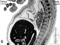
File:Human- fetal week 10 upper body B.jpg ...he embryonic period (up to week 8) but still only 2 weeks into early fetal development.(600 × 450 (105 KB)) - 15:51, 27 April 2010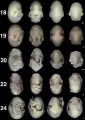
File:Bat embryo stage 18 to 24.jpg ==Bat Embryonic Development (stage 18-24)== |+ '''Embryonic Bat Stages ''Carollia perspicillata'''''<ref name="PMID15861401"><pubmed>15(800 × 1,134 (104 KB)) - 12:22, 3 July 2012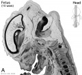
File:Human- fetal week 10 head A.jpg ...he embryonic period (up to week 8) but still only 2 weeks into early fetal development.(600 × 544 (113 KB)) - 21:22, 29 May 2011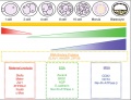
File:Mouse- preimplantation gene expression.jpg ==Characterization of global gene expression patterns during preimplantation development== ...in the characterization of gene expression patterns during preimplantation development as they have produced extensive catalogues of global stage specific gene ex(800 × 612 (106 KB)) - 09:59, 12 October 2010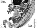
File:Human- fetal week 10 lower body C.jpg ...he embryonic period (up to week 8) but still only 2 weeks into early fetal development.(600 × 450 (94 KB)) - 15:47, 27 April 2010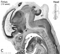
File:Human- fetal week 10 head C.jpg ...he embryonic period (up to week 8) but still only 2 weeks into early fetal development.(600 × 544 (118 KB)) - 15:47, 27 April 2010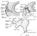
File:Rugh 128.jpg ==Development of the olfactory organ of the frog== (Bottom) Schematic reconstruction of the embryonic olfactory organ.(826 × 800 (145 KB)) - 15:40, 14 April 2013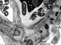
File:Fetal 10wk urogenital 1.jpg ...he embryonic period (up to week 8) but still only 2 weeks into early fetal development.(800 × 600 (109 KB)) - 21:52, 8 July 2012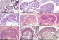
File:Human embryonic tongue 01.jpg Photographs of mid-sagittal sections of an embryonic human tongue. (A, B) Tongue development (TD) stage 1. Co, copula; Ce, foramen cecum.(1,200 × 818 (443 KB)) - 14:12, 23 March 2016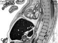
File:Human- fetal week 10 upper body C.jpg ...he embryonic period (up to week 8) but still only 2 weeks into early fetal development.(600 × 450 (109 KB)) - 15:46, 27 April 2010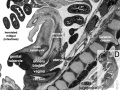
File:Fetal 10wk urogenital 4.jpg ...he embryonic period (up to week 8) but still only 2 weeks into early fetal development.(800 × 600 (105 KB)) - 17:58, 28 May 2011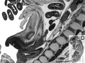
File:Human- fetal week 10 urogenital D.jpg ...he embryonic period (up to week 8) but still only 2 weeks into early fetal development.(600 × 450 (101 KB)) - 17:53, 28 May 2011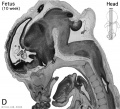
File:Human- fetal week 10 head D.jpg ...he embryonic period (up to week 8) but still only 2 weeks into early fetal development.(600 × 544 (111 KB)) - 14:50, 23 April 2013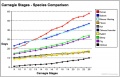
File:Carnegie stages species comparison.jpg * [[Monkey Development|Rhesus Monkey]]<ref>Hendrickx and Sawey '''Embryology of the rhesus monkey' ...Development|Mouse]]<ref>Theiler, K. '''The House Mouse: Atlas of Embryonic Development.''' 1989. New York: Springer-Verlag. ISBN 3 540 05940 7</ref>(800 × 514 (85 KB)) - 18:16, 3 June 2011
File:HeartILP001.jpg Embryo at approximately 18 days showing early angiogenesis and the development of the primordial heart tubes in the cardiogenic region. ...links to other resources. For example [[Carnegie_stage_7|Carnegie stage 7 embryonic disc]](1,507 × 898 (125 KB)) - 13:12, 8 September 2009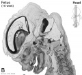
File:Human- fetal week 10 head B.jpg ...he embryonic period (up to week 8) but still only 2 weeks into early fetal development.(600 × 544 (66 KB)) - 21:22, 29 May 2011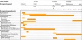
File:Human- genital development critical periods.jpg ==Human Genital Development Critical Periods== This figure provides a broad general summary of key events in genital development in relation to critical periods.(1,000 × 494 (78 KB)) - 12:12, 15 May 2019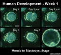
File:Human blastocyst day 1-5.jpg Human blastocyst development (in vitro) from day 1 to day 5. ...asive imaging of human embryos before embryonic genome activation predicts development to the blastocyst stage.(500 × 450 (46 KB)) - 13:50, 15 March 2014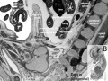
File:Fetal 10wk urogenital 2.jpg ...he embryonic period (up to week 8) but still only 2 weeks into early fetal development.(800 × 600 (110 KB)) - 21:17, 29 May 2011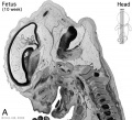
File:Human- fetal week 10 head A1.jpg ...he embryonic period (up to week 8) but still only 2 weeks into early fetal development.(1,200 × 1,088 (159 KB)) - 06:58, 7 October 2010


