Search results
From Embryology
File:Mall1917 fig02.jpg ...o. 12S9 from Dr. J. R. Cottell, Louisville, Ky. X 2. The picture shows the coelom filled mostly with granular magma.(629 × 525 (29 KB)) - 18:31, 5 November 2013
File:Wyburn1939-fig16.jpg ==Fig. 16. Photomicrograph of endothelial lining of umbilical cord coelom of embryo 42 mm== ...to cord “pillars” and continuous with rectus sheath. U.C. = umbilical cord coelom. L. = degenerating endothelial cells. Description in text.(848 × 800 (204 KB)) - 17:01, 14 September 2015
File:Wyburn1939-fig15.jpg ==Fig. 15. Photomicrograph of section of umbilical cord coelom of embryo 42 mm== ...to cord “pillars” and continuous with rectus sheath. U.C. = umbilical cord coelom. L. = degenerating endothelial cells. Description in text.(848 × 800 (189 KB)) - 17:02, 14 September 2015
File:Wyburn1939-fig13.jpg Fig. 16. Photomicrograph of endothelial lining of umbilical cord coelom of embryo 42 mm. x circa 225. ...to cord “pillars” and continuous with rectus sheath. U.C. = umbilical cord coelom. L. = degenerating endothelial cells. Description in text.(848 × 800 (153 KB)) - 17:02, 14 September 2015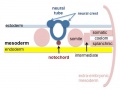
File:Mesoderm-cartoon4.jpg ...inar embryo showing the further development of the 3 layers and the space (coelom) that forms in the mesoderm (only the righthand side is shown, lefthand sid ...sion into somatic and {{splanchnic mesoderm}} separated by intra-embryonic coelom.(400 × 300 (20 KB)) - 10:09, 23 April 2020
File:Wyburn1939-plate04.jpg Fig. 15. Photomicrograph of section of umbilical cord coelom of embryo 42 mm. Site indicated by arrow in Fig. 14. x circa 75. Fig. 16. Photomicrograph of endothelial lining of umbilical cord coelom of embryo 42 mm. x circa 225.(1,700 × 2,247 (747 KB)) - 09:38, 15 September 2015
File:Wyburn1939-plate02.jpg ...I . =intercoelomic septum. D. = distal slit-like portion of umbilical cord coelom. S. = space in umbilical cord. U. = umbilical vein.(1,680 × 2,367 (786 KB)) - 17:13, 14 September 2015
File:Wyburn1939-text-fig08-10.jpg ...Horizontal lines = blood vessels. Black = mesoderm. U .C. = umbilical cord coelom. ...Horizontal lines = blood vessels. Black = mesoderm. U .C. = umbilical cord coelom.(1,588 × 1,400 (183 KB)) - 13:34, 15 September 2015
File:Dandy1910-plate02.jpg ...leural coelom; Coe, Peritoneal coelom; E.C., External Communication of the coelom: Pr, Pronephros; Ht, Projection of the heart in the pericardial cavity; X.(1,754 × 2,400 (951 KB)) - 12:10, 28 May 2017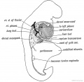
File:Keith1902 fig201.jpg ==Fig 201. The Form of the Coelom in a 3rd week Embryo as viewed from the right side==(826 × 800 (87 KB)) - 09:32, 20 January 2014
File:Bremer1914 plate05.jpg ...and mesothelial cord, leading from funnel (f.) to unlined space (a). Coe., coelom; h, smaller unlined space;c, endothelial cord. X circa 580. ...space, containing corpuscles. c and d, mesothelial cords (see text); Coe., coelom. X circa 800.(643 × 1,000 (144 KB)) - 21:50, 27 October 2015
File:Minot1889 fig04.jpg V, vein; Ar, artery; All, allantois cavity; Coe, coelom; Y, yolk sack ; X 22 diams.(1,000 × 482 (153 KB)) - 11:52, 8 May 2018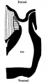
File:Wyburn1939-text-fig07.jpg .... Horizontal lines = blood vessel. Black = mesoderm. U.C. = umbilical cord coelom.(539 × 928 (46 KB)) - 13:32, 15 September 2015
File:Keith1921 fig016.jpg ==Fig. 16. Showing the Origin of the Primitive Coelom, the Mesoblast and Cavity of the Amnion during the Development of the Human(1,075 × 800 (203 KB)) - 09:57, 22 December 2014
File:Keith1902 fig073.jpg ==Fig. 73 Diagrammatic section of the abdominal region of the coelom==(800 × 577 (97 KB)) - 10:07, 7 January 2014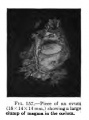
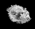
File:Keibel Mall 157.jpg Piece of an ovum (18x14x14 mm) showing a large clump of magma in the coelom.(300 × 406 (17 KB)) - 05:23, 6 September 2012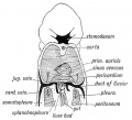
File:Keith1902 fig183.jpg ...183. Diagram to show the manner in which the Ducts of Cuvier encircle the Coelom at the junction of the Pericardial and Pleural Parts (Iter venous)==(879 × 800 (113 KB)) - 09:32, 20 January 2014
File:Wyburn1939-fig14.jpg ...to cord “pillars” and continuous with rectus sheath. U.C. = umbilical cord coelom. L. = degenerating endothelial cells. Description in text.(848 × 800 (164 KB)) - 17:02, 14 September 2015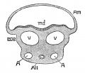
File:Minot1889 fig02.jpg ...ullary groove; v, v, veins; A, A, umbilical arteries; All, allantois; coe, coelom.(760 × 650 (91 KB)) - 11:51, 8 May 2018
File:Dandy1910-plate01.jpg '''Fig. 3.''' Section 76, X 50. Pr, Pronephros; Coe, Coelom; Pl, Pleural Cuelom; V, Umbilical vein; Br V, Branch of umbilical vein, onl ...r tip of somite IV on opposite side) ; Coe, Coelom; E.C., Communication of coelom with exterior; Ch, Chorda.(1,738 × 2,359 (541 KB)) - 12:09, 28 May 2017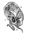
File:Mall1906-fig06.jpg (No. 12 of my collection). X 50. 03 third occipital myotome; coe, coelom; v, vein; st, septum transversum; l, liver; ph, pharynx; uv, umbilical vesi(600 × 716 (111 KB)) - 18:55, 20 September 2015
File:Stage 13 image 068.jpg ...the most cranial extension. This attachment now divides the intraembryonic coelom around the trachea into two canals, the L and R pleuro (pericardio-peritone ** Canals are lined by coelomic mesothelium and are continuous with whole I-E coelom (referred to hereafter simply as coelomic canals).(1,000 × 557 (97 KB)) - 10:00, 9 June 2011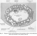
File:Hamilton1949 fig12.jpg ...Diagram to show the development of the secondary yolk sac, extra-embryonic coelom and remnant of the primary yolk sac==(1,312 × 1,265 (267 KB)) - 15:30, 15 May 2018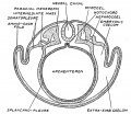
File:Keith1921 fig039.jpg ...ction across a vertebrate embryo to show the parts of the mesoderm, of the coelom==(815 × 706 (128 KB)) - 11:42, 23 December 2014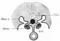
File:Bailey306.jpg * Coel - Coelom(624 × 419 (35 KB)) - 17:51, 19 September 2011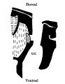
File:Wyburn1939-text-fig06.jpg ...gut. Oblique line = blood vessels. Black = mesoderm. U.C. =umbilical cord coelom.(579 × 750 (42 KB)) - 13:25, 15 September 2015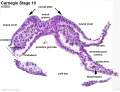
File:Stage10 K12202-04.jpg ...e 3 germ cell layers and the lateral plate divided into the intraembryonic coelom and associated mesoderm developmental regions. See [[:File:Stage10 K12202-0(1,434 × 1,105 (227 KB)) - 14:47, 15 August 2016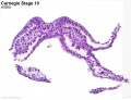
File:Stage10 K12202-03.jpg ...e 3 germ cell layers and the lateral plate divided into the intraembryonic coelom and associated mesoderm developmental regions. See [[:File:Stage10 K12202-0(1,434 × 1,105 (191 KB)) - 14:46, 15 August 2016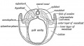
File:Keith1902 fig069.jpg ...otochord, (3) the" ingrowth of the mesoblast, and (4) the formation of the coelom.(1,000 × 548 (93 KB)) - 09:57, 7 January 2014
File:1901 Comparative development of the coelom.pdf ==Comparative Development of the Coelom==(5.49 MB) - 10:49, 20 February 2020
File:Bailey052.jpg The parietal mesoderm (lying above the coelom) is not labeled.(895 × 515 (99 KB)) - 23:41, 13 February 2011
File:Bailey101.jpg The parietal mesoderm (lying above the coelom) is not labeled.(900 × 533 (110 KB)) - 12:45, 18 January 2011
File:Dandy1910-plate06.jpg ...All - Allantois Ch - Chorda; Ht - Heart; Coe - Coelom; P.C. — Pericardial coelom; Ca - Capillary, but no connection; Pl — Plexus of lateral aortic branche(1,000 × 2,166 (265 KB)) - 12:12, 28 May 2017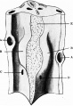
File:Waterston1915 fig04.jpg ==Fig. 4. Dorsal wall of the pericardial and upper peritoneal portions of coelom with pericardio-peritoneal passages and commencing lung buds==(633 × 919 (123 KB)) - 16:56, 24 August 2015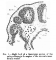
File:Low 01.jpg * Coe., coelom(502 × 535 (60 KB)) - 02:44, 29 May 2017
File:Minot1897 fig053.jpg Am.c, Amniotic cavity. Coe, Coelom. Ec, Ectoderm, in B, bearing the anlages of villi. Ent, Entoderm.(1,363 × 631 (94 KB)) - 17:11, 27 July 2015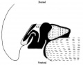
File:Wyburn1939-text-fig04.jpg ...liver. small dots = gut and allantoic diverticulum. U.C. = umbilical cord coelom. Black line indicates the plane of the umbilical veins.(1,117 × 882 (83 KB)) - 13:12, 15 September 2015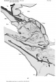
File:Bremer1914 plate01.jpg ...nections with the mesothelium. All., allantois; bl.i.. blood-island; Coe., coelom; Ect., ectoderm of chorion; f. , funnel-shaped connections, with unconnecte(684 × 1,000 (102 KB)) - 14:35, 12 August 2019
File:Odgers1937 plate02fig03.jpg ...he yolk sac surrounded on either side and ventrally by the extra-embryonic coelom.(1,000 × 692 (167 KB)) - 21:24, 29 June 2015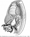
File:Bailey266.jpg The intestinal coils lie for the most part in the umbilical coelom. ...e and form secondary loops, all of which push their way into the umbilical coelom where they remain until the embryo reaches a length of 40 mm (compare [[Boo(731 × 913 (178 KB)) - 16:31, 15 April 2014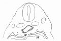
File:Flint1906 textfig01.jpg C = Coelom. SV = Sinus venosus. VM = Mesocardium posterior.(700 × 473 (33 KB)) - 14:21, 8 April 2020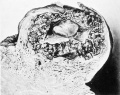
File:Mall1916 fig03.jpg The embryo lies within the coelom, and bands of -magma fibrils radiate from the amnion to the chorionic wall.(995 × 787 (145 KB)) - 10:51, 22 April 2014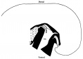
File:Wyburn1939-text-fig05.jpg ...blood vessels. Black = mesoderm. Small dots = gut. U .C. = umbilical cord coelom. Black line indicates the plane of the umbilical veins.(1,300 × 935 (66 KB)) - 13:19, 15 September 2015
File:Minot1889 fig01.jpg ...doderm only; vt, vena terminals; mrs, mesoderm; sol, splanchnopleure; coe, coelom; nch, notochord; Md, medullary groove; my, myotome; Endo, endoderm; Ecto, e(1,200 × 644 (307 KB)) - 12:17, 4 April 2014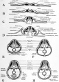
File:Patten054.jpg ...grams of cross sections at various stages to show the establishment of the coelom and mesenteries== ...all in the yolk-stalk region, results in the embryonic and extra-embryonic coelom retaining their open communication at this point for a long time after they(764 × 1,060 (179 KB)) - 09:11, 29 July 2011
File:HHstage8-.jpg | 1-3 somites; coelom(727 × 1,125 (90 KB)) - 16:22, 2 June 2017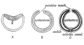
File:Keith1921 fig038.jpg '''C.''' Origin of mesoderm (black) and coelom from margin of primitive mouth, with formation of a ventral mesentery round(1,200 × 636 (118 KB)) - 11:40, 23 December 2014
File:Hertig1946b fig09b.jpg ...he communication between the exocoelomic space around the yolk-sac and the coelom (body cavity) within the embryo. Carnegie {{CE6488}}, sequence 2, X15. 104(800 × 586 (26 KB)) - 17:35, 7 August 2017
File:Wyburn1939-plate01.jpg A. = amnion. L.T. = lateral tissue plate. U. = umbilical vein. C. = coelom. Description in text.(1,600 × 2,180 (696 KB)) - 17:17, 14 September 2015
File:Wyburn1937 text-fig10.jpg ...cranial attachment of body stalk; between B and C is the extension of the coelom occupied by the midgut loop; D = the caudal attachment of the stalk; G.t. =(803 × 620 (40 KB)) - 17:53, 15 August 2015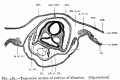
File:Bailey483.jpg ...nt at which extraembryonic body cavity passes over into intraembryonic (or coelom).(753 × 499 (64 KB)) - 08:47, 3 February 2011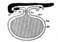
File:Bailey485.jpg ...f yolk sac; hs., parietal layer of yolk sac; hn., dermal umbilicus; lh l , coelom; lh 2 , exoccelom; m., mouth; st., yolk stalk.(583 × 425 (57 KB)) - 08:57, 3 February 2011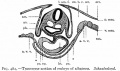
File:Bailey482.jpg ...t which extraembryonic body cavity passes over into the intraembryonic (or coelom proper).(709 × 419 (57 KB)) - 16:23, 9 August 2012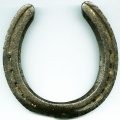
File:Horseshoe.jpg ...to describe the shape of the [[Coelomic Cavity Development|intra-embryonic coelom]] within the lateral plate [[mesoderm]] that forms during week 3 of human d(400 × 400 (32 KB)) - 09:09, 13 October 2016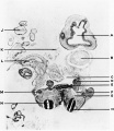
File:Shaw1932 fig03.jpg A, cerebral vesicle; B, heart; 0, coelom; D, intestine; E, primitive kidney; F, limb bud; G, dorsal aorta; H, neural(900 × 1,041 (81 KB)) - 17:09, 15 November 2017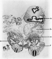
File:Shaw1932 fig04.jpg A, cerebral vesicle; B, heart; 0, coelom; D, intestine; E, primitive kidney; F, limb bud; G, dorsal aorta; H, neural(900 × 1,021 (100 KB)) - 17:09, 15 November 2017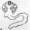
File:Hertig1946b fig10b.jpg ...of the embryo. The large spaces on either side of the gut are those of the coelom or body-cavity of the embryo. Carnegie {{CE6344}}, section 3-7-11, X75.(800 × 818 (85 KB)) - 08:37, 8 August 2017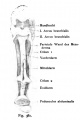
File:Kollmann381.jpg The intestinal system still consists of a straight tube of the Coelom surrounding the elongated cylindrical body, heart and runs Yolk sac are rem(463 × 695 (31 KB)) - 12:07, 20 October 2011
File:Flint1906 textfig02.jpg Through the pulmonary anlage. C = Coelom. PA = Pulmonary anlage.(671 × 520 (37 KB)) - 13:35, 8 April 2020
File:Wyburn1937 text-fig06.jpg ...ion 910; C = section 954; interval between B and 0 is the extension of the coelom into the stalk and occupied by the midgut loop. D = section 1002 at the lev(1,021 × 528 (55 KB)) - 17:44, 15 August 2015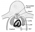
File:Rugh 156.jpg ==The coelom and its derivatives in the frog==(700 × 655 (86 KB)) - 22:12, 27 September 2013
File:Waterston11.jpg * '''E''' - coelom(500 × 728 (75 KB)) - 12:34, 28 January 2012
File:Keith1921 fig021.jpg | 6. Coelom, bounded by the somatopleure externally and splanchnopleure internally.(1,179 × 625 (160 KB)) - 10:13, 22 December 2014
File:Stage 13 image 085.jpg Note rectum, urogenital sinus, allantois and intraembryonic coelom.(1,000 × 434 (99 KB)) - 15:32, 21 September 2010
File:Stage 13 image 084.jpg Note shape of Intraembryonic coelom on L. Site of caudal attachment of connecting stalk to body wall.(1,000 × 420 (101 KB)) - 15:35, 21 September 2010
File:Waterston1915 fig05.jpg ...ricardial sac, pericsrdio-peritoneal passages and upper part of peritoneal coelom in 6 mm Embryo==(898 × 559 (108 KB)) - 17:07, 24 August 2015
File:DeBeer1928 fig127.jpg Showing the relations of the coelom and viscera. a, anus ; au, auricle of heart ; b, bladder (continuous with a(1,055 × 1,284 (288 KB)) - 17:30, 26 April 2015
File:Stage13 bf5.jpg * Larger cavity is chorionic space (coelom).(901 × 676 (65 KB)) - 08:30, 28 August 2013
File:Stage13 bf6.jpg * Larger cavity is chorionic space (coelom).(901 × 676 (80 KB)) - 14:49, 28 February 2014
File:Stage13 bf6.gif * Larger cavity is chorionic space (coelom).(901 × 676 (463 KB)) - 08:33, 28 August 2013
File:Wyburn1937 text-fig08.jpg ...the body stalk. B =section 1200. 0 = section 1256. B-C = extension of the coelom occupied by the midgut loop. D = section 1272 at the level of the caudal at(1,417 × 624 (114 KB)) - 17:49, 15 August 2015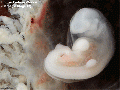
File:Stage13 bf5.gif * Larger cavity is chorionic space (coelom).(600 × 450 (197 KB)) - 08:24, 12 August 2011
File:Wyburn1937 text-fig07.jpg ...on 972; 0 = section 1044; interval between B and 0 is the extension of the coelom occupied by the midgut loop. D = section 1052 at the level of caudal attach(1,143 × 802 (107 KB)) - 17:58, 15 August 2015
File:Wyburn1937 text-fig03.jpg ...al between B and 0 is the communication between extra- and intra-embryonic coelom. D = the level of the caudal attachment of the body stalk.(1,629 × 700 (160 KB)) - 18:00, 15 August 2015
File:Wyburn1937 text-fig11.jpg ...stalk; B = section 1184; 0 = section 1236; between B and 0 is extension of coelom; D = section 1252 at the level of the caudal attachment of stalk; G.t. = se(1,427 × 1,101 (148 KB)) - 17:55, 15 August 2015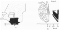
File:Wyburn1937 text-fig09.jpg ...B = section 857 ; 0 =section 913. Between B and 0 is the extension of the coelom occupied by the midgut loop. D=section 921 at the level of the caudal attac(1,276 × 638 (100 KB)) - 17:52, 15 August 2015
File:Waterston20.jpg ...septum transversum, showing the dorsal wall of pericardial and peritoneal coelom with orifices of right and left pneumato-enteric recesses.(500 × 720 (75 KB)) - 21:57, 20 February 2012
File:Stage6 bf03.jpg Extra-embryonic coelom(646 × 800 (65 KB)) - 14:48, 1 October 2018
File:Kollmann061.jpg ...inal tube and its appendages throughout thevertebrate kingdom absorbs. The coelom does not extend into the root zone (Fig.Gl), and not in the front section o(1,000 × 512 (78 KB)) - 16:24, 30 October 2011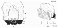
File:Wyburn1937 text-fig05.jpg ...ion 775; C = section 825; interval between B and C is the extension of the coelom into the stalk and occupied by mid-gut loop; D = section 875 at the level o(1,122 × 545 (85 KB)) - 17:59, 15 August 2015
File:Stage14 bf27.jpg * Larger cavity is chorionic space (coelom).(1,000 × 1,291 (208 KB)) - 22:07, 14 June 2016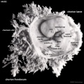
File:HillH202 Stage 17 bf04.jpg * chorionic cavity - The fluid-filled extraembryonic coelom (cavity) formed initially from trophoblast and extraembryonic mesoderm that * '''amniotic cavity''' - The fluid-filled (amniotic fluid) extraembryonic coelom (cavity) formed initially by epiblast and then lined by ectoderm and surrou(1,000 × 1,000 (138 KB)) - 17:58, 22 March 2014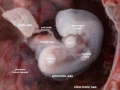
File:Stage14 bf24.jpg * Larger cavity is chorionic space (coelom).(1,231 × 923 (156 KB)) - 06:54, 7 April 2016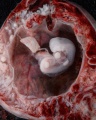
File:Stage14 bf20.jpg * Larger cavity is chorionic space (coelom).(800 × 1,000 (158 KB)) - 14:43, 28 February 2014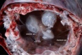
File:Stage14 bf25.jpg * Larger cavity is chorionic space (coelom).(2,000 × 1,334 (337 KB)) - 16:02, 16 March 2014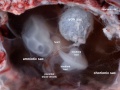
File:Stage14 bf26.jpg * Larger cavity is chorionic space (coelom).(1,263 × 947 (150 KB)) - 16:02, 16 March 2014
File:Stage14 bf19.jpg * Larger cavity is chorionic space (coelom).(1,200 × 1,500 (287 KB)) - 14:44, 28 February 2014
File:Bardeen1906-plate01.jpg ...left the bud is shown cut through an area near the distal extremity of the coelom. At the right the cut is more dorsal and extends through the tips of the lu(1,565 × 2,322 (238 KB)) - 23:06, 23 July 2020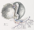
File:Cullen1916 fig02.jpg enveloping the embryo. Compare the situation of the coelom in this with that in the subsequent pictures. There is(1,280 × 1,110 (403 KB)) - 09:21, 28 October 2018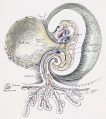
File:Cullen1916 fig03.jpg ...The exocoelom has been drawn into the embryo and will later unite with the coelom of the pleuroperitoneal cavity.(1,280 × 1,443 (494 KB)) - 10:01, 30 October 2018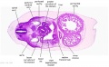
File:Stage 13 image 073.jpg * Thick mesothelium lining the coelom along L edge of stomach, the primordium of the spleen and greater omentum a(1,000 × 619 (118 KB)) - 07:37, 15 May 2014
File:Shaw1932 fig02.jpg Figs. 3 and 4. A, cerebral vesicle; B, heart; 0, coelom; D, intestine; E, primitive kidney; F, limb bud; G, dorsal aorta; H, neural(1,000 × 653 (49 KB)) - 17:08, 15 November 2017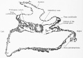
File:Keibel Mall 2 525.jpg ...mitive segment stalk, remaining narrow. On the left side of the figure the coelom of the primitive segment stalk and that of the lateral plate are separate,(1,280 × 896 (160 KB)) - 16:45, 14 November 2018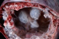
File:Stage14 bf22.jpg * Larger cavity is chorionic space (coelom).(1,500 × 1,001 (214 KB)) - 14:48, 28 February 2014
File:Stage14 bf23.jpg * Larger cavity is chorionic space (coelom).(1,000 × 667 (122 KB)) - 14:48, 28 February 2014
File:Stage14 bf21.jpg * Larger cavity is chorionic space (coelom).(2,000 × 1,334 (323 KB)) - 14:49, 28 February 2014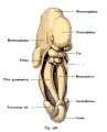
File:Kollmann426.jpg The heart and pulmonary system remained. The dorsal half shows the coelom and the two primitive kidneys (Mesonephroi). Over the anterior surface(500 × 620 (46 KB)) - 12:22, 20 October 2011
File:Hertig1946b fig10.jpg ...of the embryo. The large spaces on either side of the gut are those of the coelom or body-cavity of the embryo. Carnegie {{CE6344}}, section 3-7-11, X75.(800 × 1,341 (118 KB)) - 08:34, 8 August 2017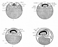
File:Bailey484.jpg al., Allantois; am., amnion; am. c., amniotic cavity; cce., coelom; dh., vitelline area between two dotted lines which represent the edge of t(869 × 707 (119 KB)) - 16:27, 9 August 2012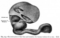
File:Bailey264.jpg ...e and form secondary loops, all of which push their way into the umbilical coelom where they remain until the embryo reaches a length of 40 mm. (compare Figs(881 × 562 (77 KB)) - 12:27, 15 April 2011


