Uploads by Z3212774
From Embryology
This special page shows all uploaded files.
| Date | Name | Thumbnail | Size | Description | Versions |
|---|---|---|---|---|---|
| 11:28, 14 March 2010 | Ventricular Septal Defect.jpg (file) |  |
16 KB | category:Heart ILP | 1 |
| 11:25, 14 March 2010 | Partial Anomalous Pulmonary Venous Drainage.jpg (file) |  |
16 KB | category:Heart ILP | 1 |
| 11:23, 14 March 2010 | Double Outlet Right Ventricle.jpg (file) |  |
16 KB | category:Heart ILP | 1 |
| 11:27, 14 March 2010 | Tricuspid Atresia.jpg (file) |  |
16 KB | category:Heart ILP | 1 |
| 11:26, 14 March 2010 | Pulmonary Stenosis.jpg (file) |  |
16 KB | category:Heart ILP | 1 |
| 11:22, 14 March 2010 | Atrial Septal Defect.jpg (file) |  |
16 KB | category:Heart ILP | 1 |
| 11:22, 14 March 2010 | Aortic Stenosis.jpg (file) |  |
16 KB | category:Heart ILP | 1 |
| 11:25, 14 March 2010 | Patent Ductus Arteriosus.jpg (file) |  |
16 KB | category:Heart ILP | 1 |
| 11:23, 14 March 2010 | Coarctation of the Aorta.jpg (file) |  |
16 KB | category:Heart ILP | 1 |
| 11:09, 29 September 2009 | HeartILP draft coarctationoftheaorta.jpg (file) |  |
16 KB | category:HeartILP | 1 |
| 11:12, 29 September 2009 | HeartILP draft interruptaorticarch.jpg (file) |  |
17 KB | category:HeartILP | 1 |
| 11:24, 14 March 2010 | Interrupted Aortic Arch.jpg (file) |  |
17 KB | category:Heart ILP | 1 |
| 11:25, 14 March 2010 | Pulmonary Atresia.jpg (file) |  |
17 KB | category:Heart ILP | 1 |
| 11:27, 14 March 2010 | Tetralogy of Fallot.jpg (file) |  |
17 KB | category:Heart ILP | 1 |
| 11:24, 14 March 2010 | Hypoplastic Left Heart.jpg (file) |  |
17 KB | category:Heart ILP | 1 |
| 11:26, 14 March 2010 | Total Anomalous Pulmonary Venous Connection.jpg (file) |  |
17 KB | category:Heart ILP | 1 |
| 11:27, 14 March 2010 | Transposition of the Great Vessels.jpg (file) |  |
18 KB | category:Heart ILP | 1 |
| 11:24, 14 March 2010 | Functional Hypoplastic Left Heart.jpg (file) |  |
18 KB | category:Heart ILP | 1 |
| 11:11, 29 September 2009 | HeartILP draft funchlh.jpg (file) |  |
18 KB | category:HeartILP | 1 |
| 09:53, 14 March 2010 | Navigation bar 2.jpg (file) | 26 KB | Heart ILP | 1 | |
| 11:39, 14 March 2010 | Aortic Arches (Drawing).jpg (file) | .jpg) |
29 KB | category:Heart ILP | 1 |
| 09:52, 14 March 2010 | Navigation bar 1.jpg (file) | 31 KB | Heart ILP | 1 | |
| 11:24, 22 September 2009 | HeartILP draft animpic005.jpg (file) |  |
49 KB | Craniocaudal patterning of cardiogenic progenitors in the epiblast and primitive streak; these migrate through the primitive streak to be organised mediolaterally in the cardiogenic mesoderm. category:HeartILP | 1 |
| 08:35, 15 October 2009 | HeartILP draft Navigation2.jpg (file) | 52 KB | 1 | ||
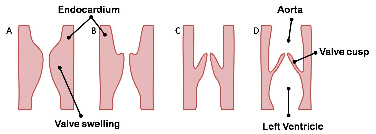
| 13:17, 2 October 2009 | HeartILP draft semilunarlongitudinal.jpg (file) |  |
52 KB | Longitudinal sections of the aorta showing development of the semilunar cusps. category:HeartILP | 1 |
| 11:17, 14 March 2010 | Semilunar Cusps.jpg (file) |  |
52 KB | category:Heart ILP Longitudinal sections of the aorta showing development of the semilunar cusps forming the aortic valve. | 1 |
| 21:59, 28 September 2009 | HeartILP draft animpic010.jpg (file) |  |
59 KB | Animation of folding of the embryo resulting in fusion of the endocardial heart tubes. category:HeartILP | 1 |
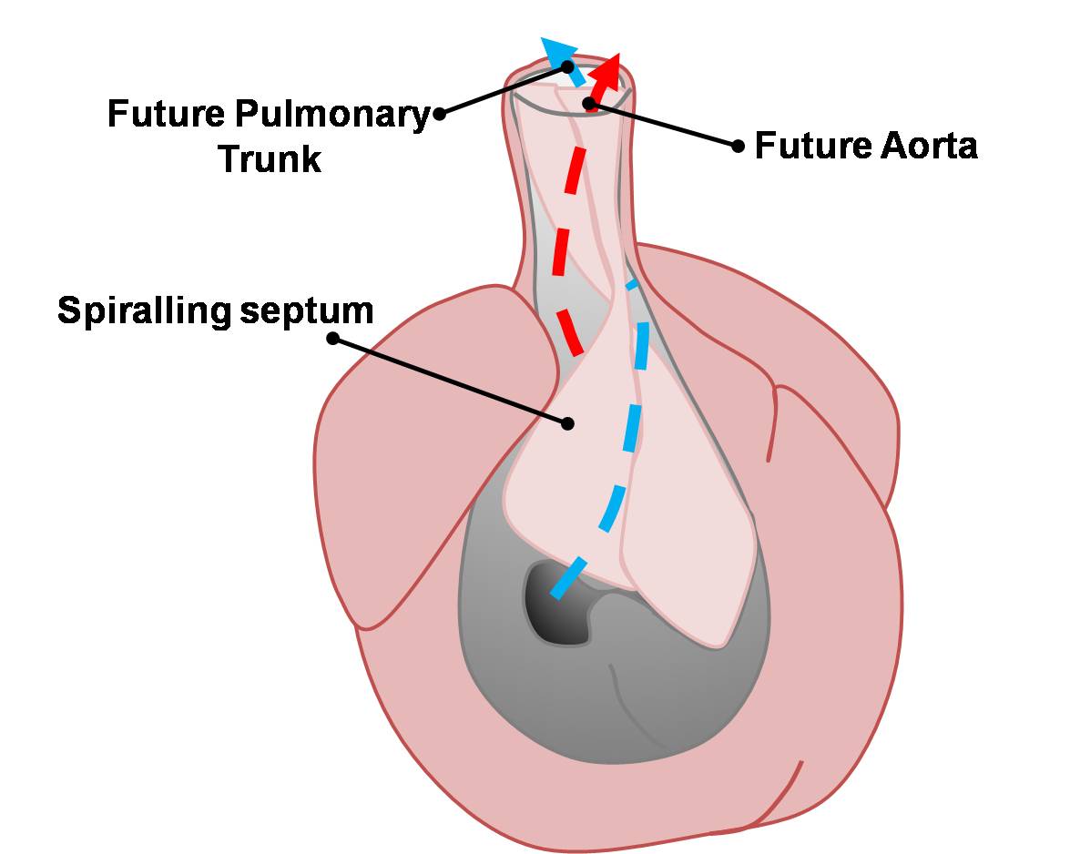
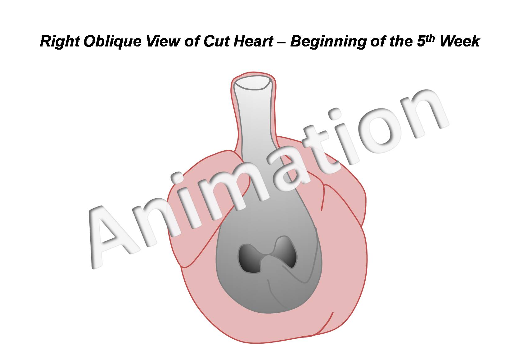
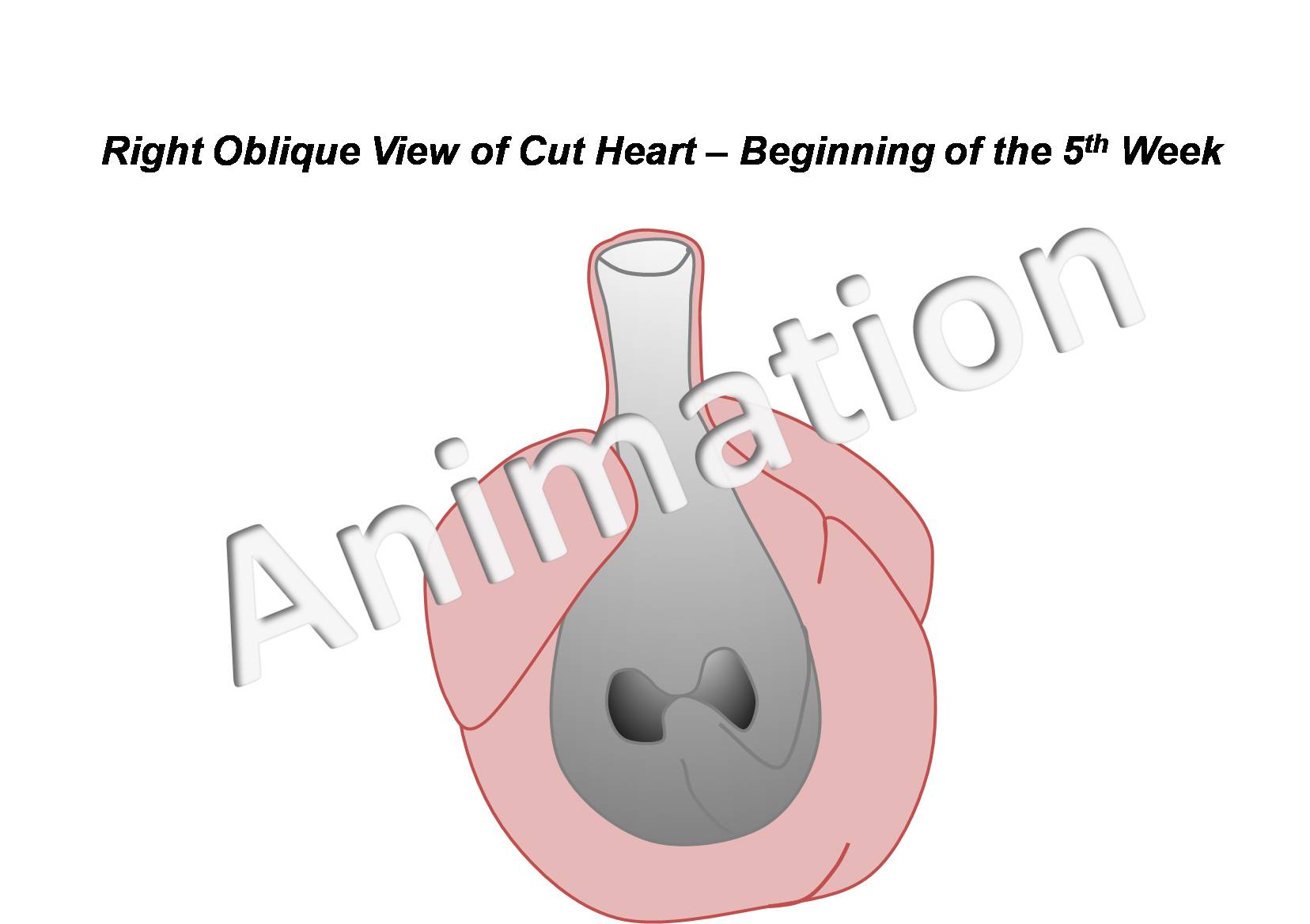
| 10:22, 14 March 2010 | Basic Outflow Tract Division.jpg (file) |  |
59 KB | category:Heart ILP Blood flow through the conotruncus is divided to form the pulmonary trunk and aorta. | 1 |
| 09:10, 29 September 2009 | HeartILP draft outflowtract2.jpg (file) |  |
59 KB | Blood flow through the conotruncus is divided to form the pulmonary trunk and aorta. category:HeartILP | 1 |
| 11:46, 8 September 2009 | HeartILP002.jpg (file) |  |
60 KB | Endocardial tubes fuse together to form the primordial heart tube. category:HeartILP | 1 |
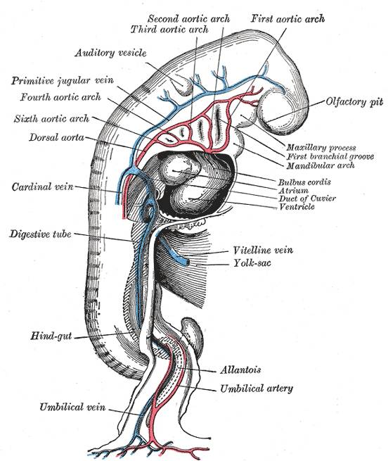
| 11:39, 14 March 2010 | Embryonic Cardiovascular System (Drawing).jpg (file) | .jpg) |
60 KB | category:Heart ILP | 1 |
| 11:00, 29 September 2009 | HeartILP draft grayembryoniccirc.jpg (file) |  |
60 KB | category:HeartILP | 1 |
| 10:15, 22 September 2009 | HeartILP draft hearttubesegments.jpg (file) |  |
63 KB | As the tubular heart grows it develops dilations and constrictions which form the truncus arteriosus, bulbus cordis, primitive ventricle, primitive atrium and sinus venosus. {{Template:SEM}} category:HeartILP | 1 |
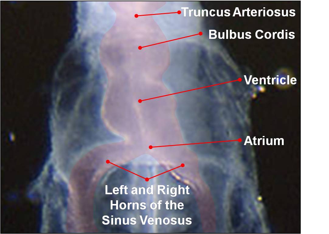
| 10:42, 14 March 2010 | Heart Tube Segments.jpg (file) |  |
63 KB | category:Heart ILP {{Template:SEM}} As the tubular heart grows it develops dilations and constrictions which form the truncus arteriosus, bulbus cordis, primitive ventricle, primitive atrium and sinus venosus. | 1 |
| 20:51, 13 October 2009 | HeartILP draft navigation.jpg (file) |  |
69 KB | 1 | |
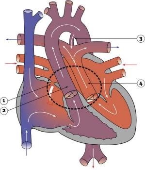
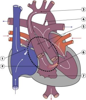
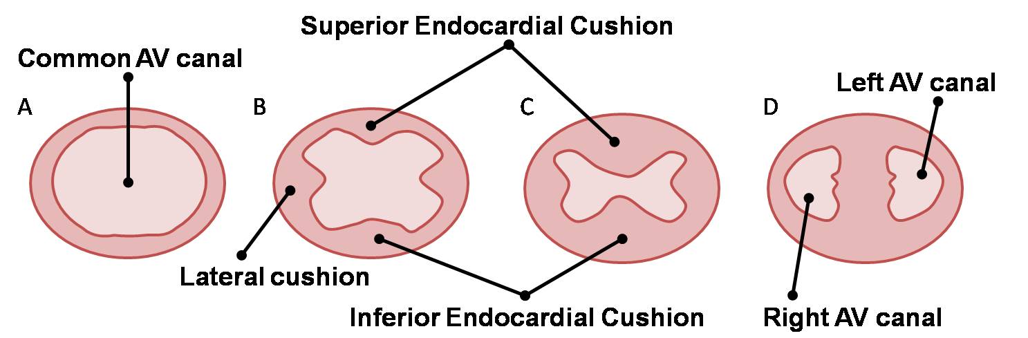
| 11:14, 14 March 2010 | AV Canal Division (Superior View).jpg (file) | .jpg) |
69 KB | category:Heart ILP Development of the atrioventricular septum over weeks four and five. The right and left atrioventricular canals are remodelled to later become the atrioventricular (tricuspid and mitral) valves. | 1 |
| 11:31, 2 October 2009 | HeartILP draft avvalves.jpg (file) |  |
69 KB | Development of the atrioventricular septum over weeks four and five. The right and left atrioventricular canals are remodelled to later become the atrioventricular (tricuspid and mitral) valves. category:HeartILP | 1 |
| 11:50, 8 September 2009 | HeartILP-draft004.jpg (file) | 70 KB | Looping of the heart tube. category:HeartILP | 1 | |
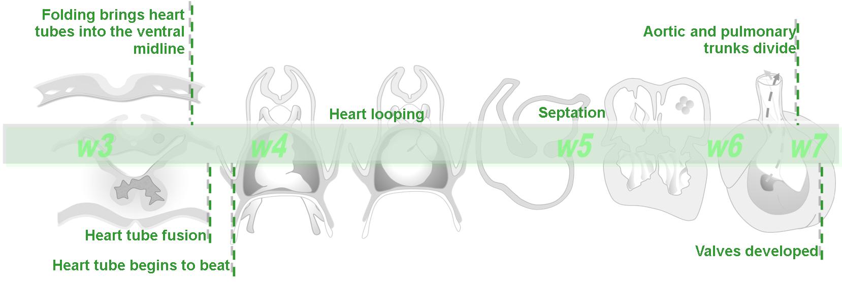
| 09:55, 14 March 2010 | Basic Heart Development Timeline.jpg (file) |  |
73 KB | category:Heart ILP | 1 |
| 11:12, 5 November 2009 | HeartILP draft btimeline.jpg (file) |  |
73 KB | 2 | |
| 11:32, 22 September 2009 | HeartILP draft animpic006.jpg (file) |  |
74 KB | Heart looping from ventral, right lateral and left lateral views with the addition of myocardium throughout looping and intrinsic and extrinsic forces controlling looping shown. category:HeartILP | 1 |
| 09:42, 14 March 2010 | Cardiac Embryology ILP Watermark.jpg (file) |  |
78 KB | category:Heart ILP | 1 |
| 20:43, 13 October 2009 | HeartILP draft heartwatermark.jpg (file) |  |
78 KB | 1 | |
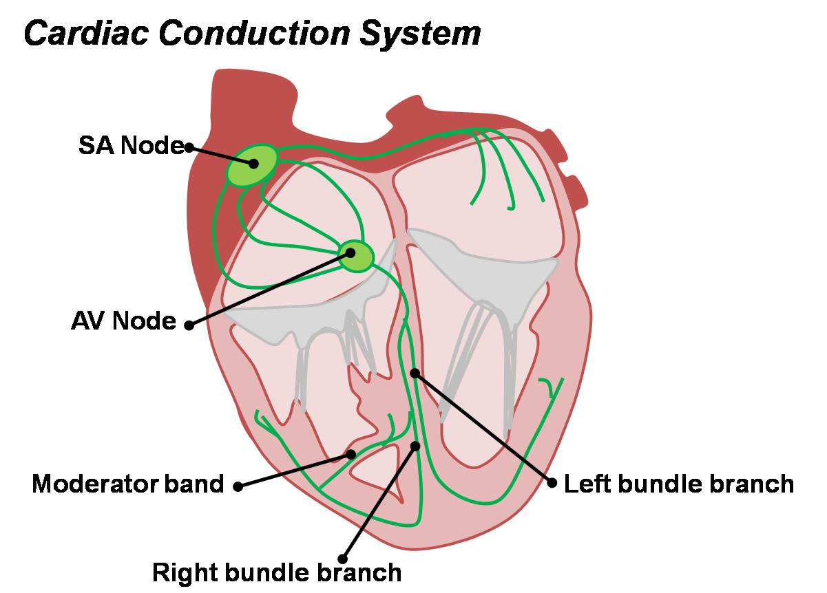
| 12:03, 14 March 2010 | Cardiac Conduction System.jpg (file) |  |
81 KB | category:Heart ILP Cardiac conduction system in the adult heart. | 1 |
| 22:04, 28 September 2009 | HeartILP draft animpic012.jpg (file) |  |
82 KB | Animation of division of the outflow tract. category:HeartILP | 1 |
| 22:03, 28 September 2009 | HeartILP draft animpic011.jpg (file) |  |
82 KB | Animation of division of the outflow tract. category:HeartILP | 1 |
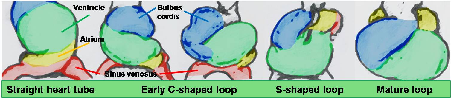
| 16:29, 29 October 2009 | HeartILP draft loopingseries.jpg (file) |  |
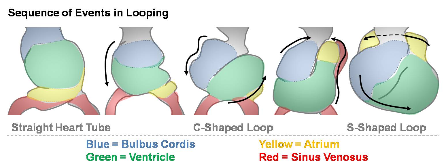
85 KB | Shows the sequence of events in heart looping. The heart begins as a straight tube then bends ventrally. Rotation brings the bulge of the ventral bend (predominantly the bulbus cordis and ventricle) to the right, forming a C-shaped loop. As the atrium | 1 |
| 10:49, 14 March 2010 | Heart Looping Sequence.jpg (file) |  |
85 KB | category:Heart ILP Shows the sequence of events in heart looping. The heart begins as a straight tube then bends ventrally. Rotation brings the bulge of the ventral bend (predominantly the bulbus cordis and ventricle) to the right, forming a C-shaped | 1 |
| 10:10, 14 March 2010 | Divisions of Early Heart Tube.jpg (file) |  |
85 KB | {{Template:SEM}} category:Heart ILP | 1 |
| 11:48, 8 September 2009 | HeartILP-draft003.jpg (file) |  |
85 KB | Early divisions of the heart tube. category:HeartILP | 1 |