Uploads by Z3212774
From Embryology
This special page shows all uploaded files.
| Date | Name | Thumbnail | Size | Description | Versions |
|---|---|---|---|---|---|
| 10:18, 14 March 2010 | AV Canal Division.jpg (file) |  |
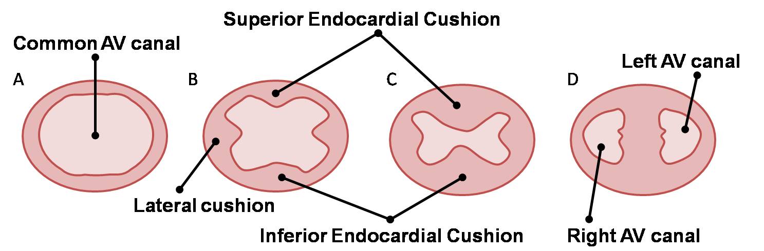
93 KB | category:Heart ILP Division of the atrioventricular canal occurs via growth and fusion of the dorsal and ventral (or superior and inferior) endocardial cushions. | 1 |
| 11:14, 14 March 2010 | AV Canal Division (Superior View).jpg (file) | .jpg) |
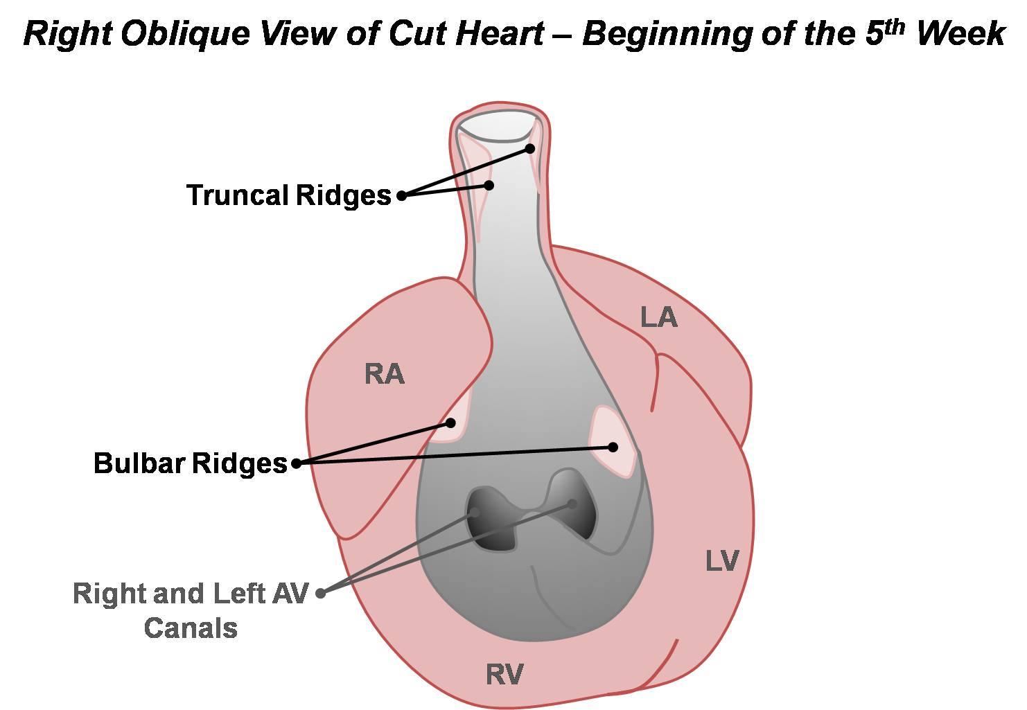
69 KB | category:Heart ILP Development of the atrioventricular septum over weeks four and five. The right and left atrioventricular canals are remodelled to later become the atrioventricular (tricuspid and mitral) valves. | 1 |
| 11:15, 14 March 2010 | AV Valves.jpg (file) |  |
129 KB | category:Heart ILP Sequence of events in the development of the atrioventricular valves. The structures of the valves i.e. the papillary muscles, chordae tendineae and cusps are sculpted from the muscular ventricular walls. | 1 |
| 11:13, 14 March 2010 | Adult Heart Valves.jpg (file) |  |
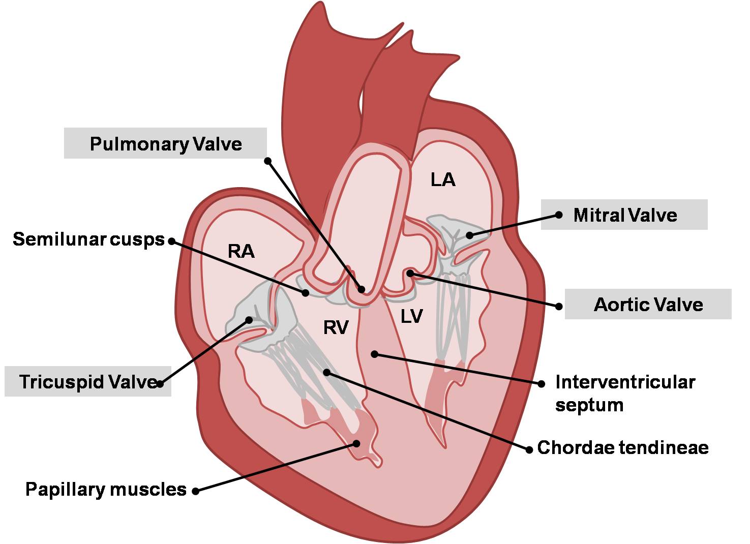
113 KB | category:Heart ILP Adult heart showing aortic, pulmonary, mitral and tricuspid valves. | 1 |
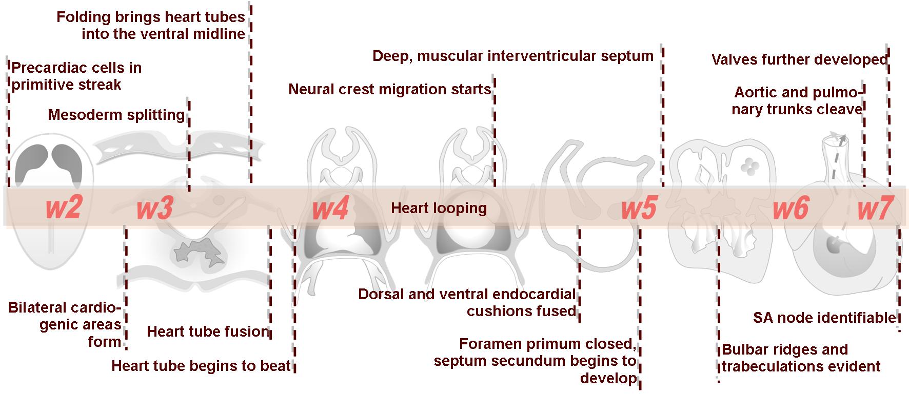
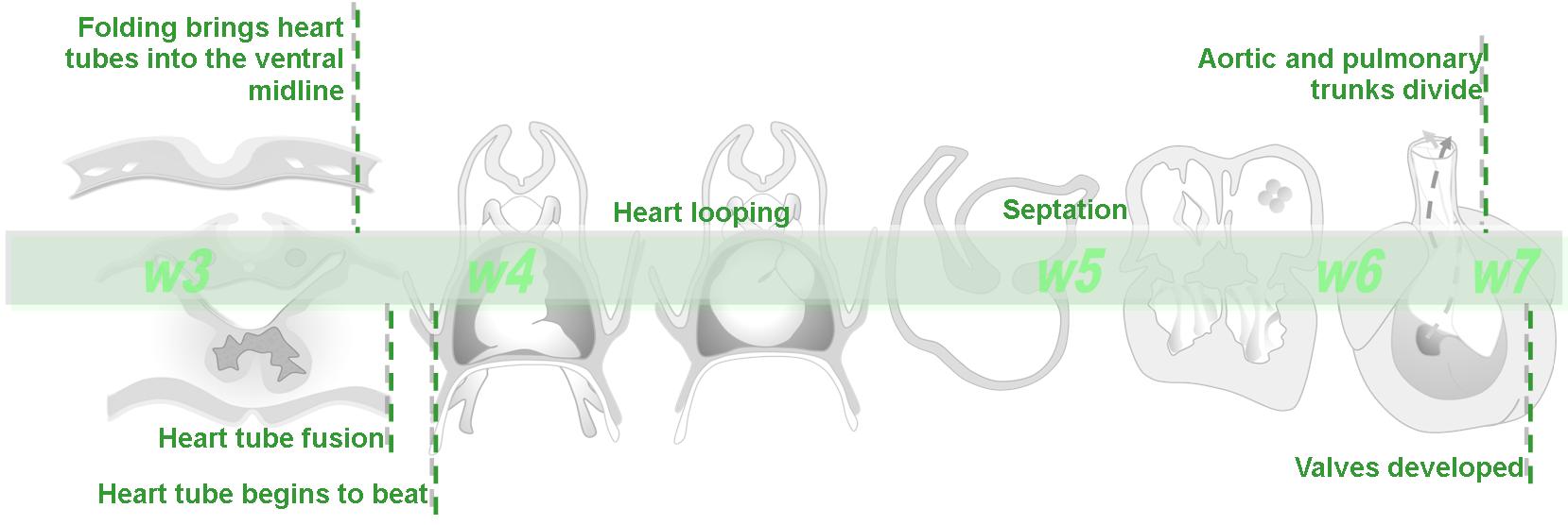
| 09:57, 14 March 2010 | Advanced Heart Development Timeline.jpg (file) |  |
158 KB | category:Heart ILP | 1 |
| 11:39, 14 March 2010 | Aortic Arches (Drawing).jpg (file) | .jpg) |
29 KB | category:Heart ILP | 1 |
| 11:22, 14 March 2010 | Aortic Stenosis.jpg (file) |  |
16 KB | category:Heart ILP | 1 |
| 10:20, 14 March 2010 | Atrial & Ventricular Septation 1.jpg (file) |  |
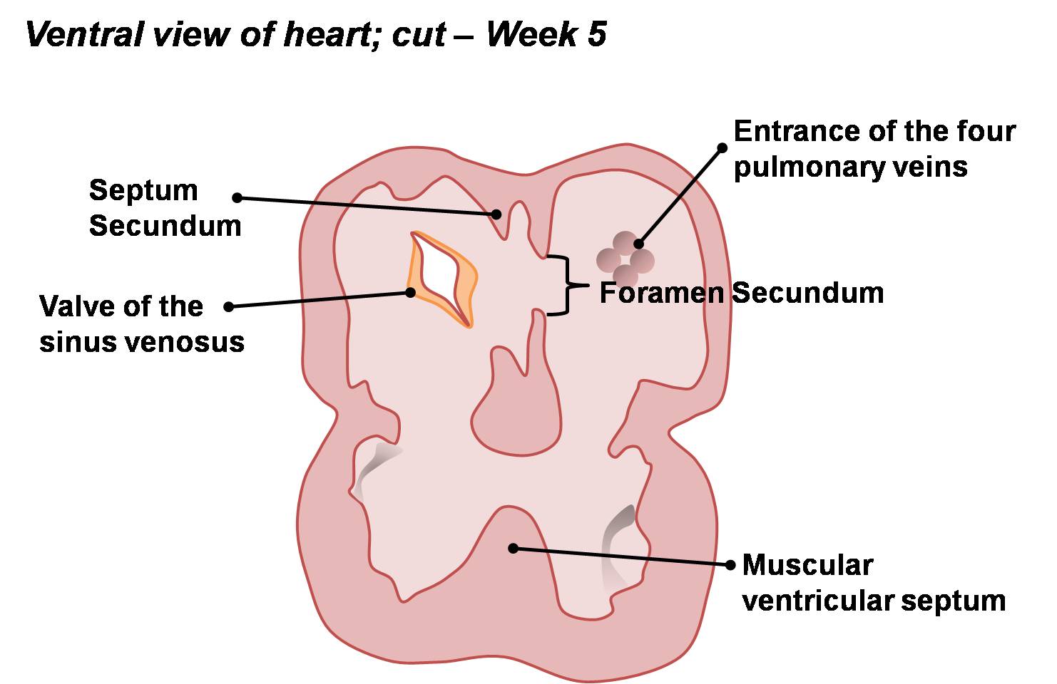
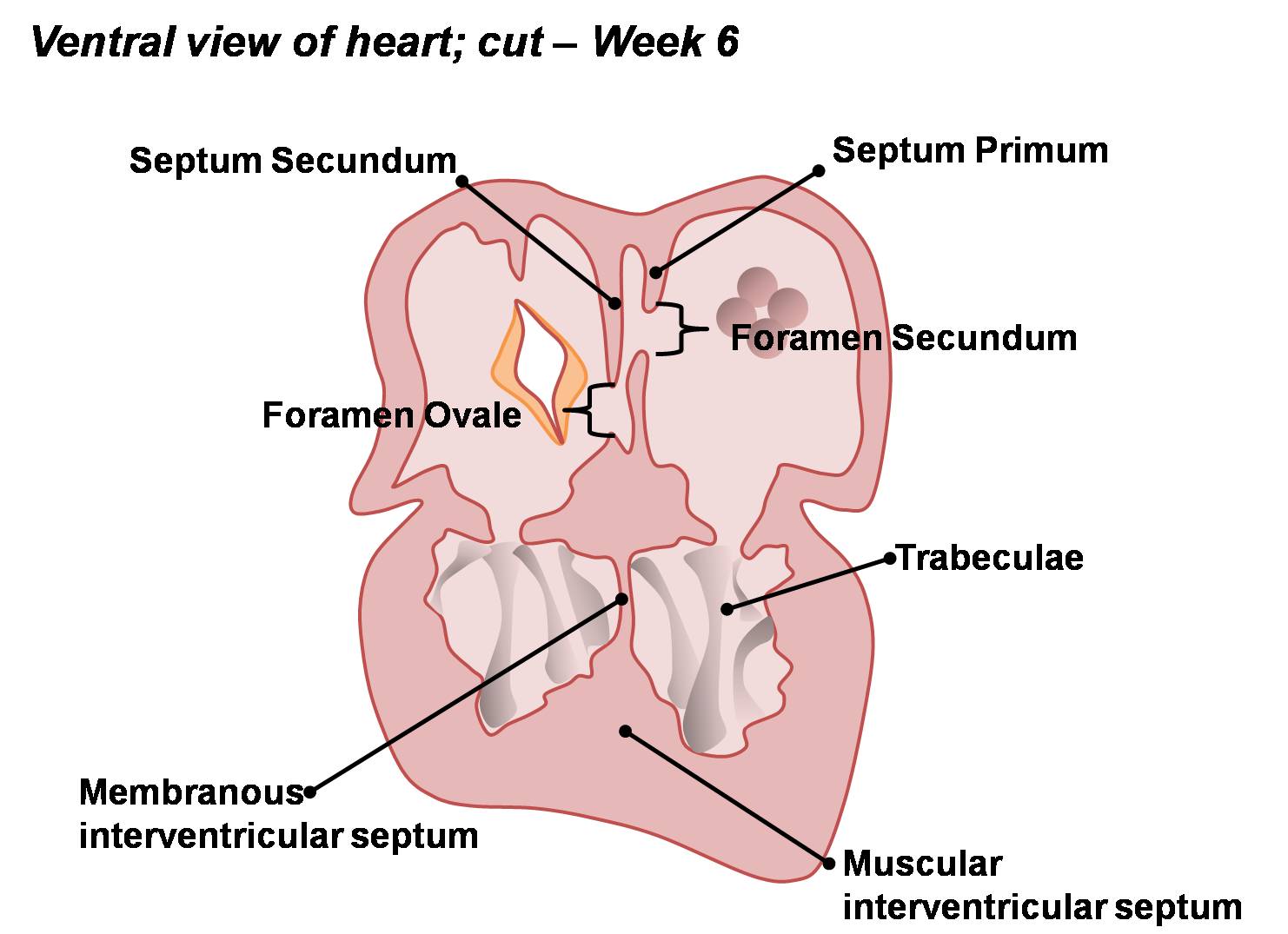
97 KB | category:Heart ILP Apoptotic induced perforations appear in the fused septum primum and form the foramen secundum. The septum secundum also begins to grow on the right of the septum primum. The muscular part of the interventricular septum develops co | 1 |
| 10:20, 14 March 2010 | Atrial & Ventricular Septation 2.jpg (file) |  |
113 KB | category:Heart ILP The embryonic heart begins to resemble the adult heart as septation is complete. A right-to-left shunt exists between the right and left atria via the foramen ovale. Both muscular and membranous portions of the ventricular septum a | 1 |
| 11:22, 14 March 2010 | Atrial Septal Defect.jpg (file) |  |
16 KB | category:Heart ILP | 1 |
| 10:19, 14 March 2010 | Atrial Septation.jpg (file) |  |
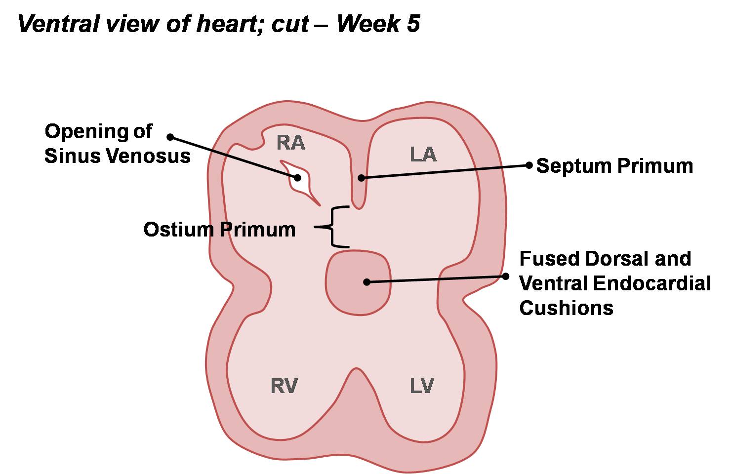
90 KB | category:Heart ILP During septation, the septum primum develops in the roof of the atrium, forming the early left and right atria. The opening of the sinus venosus has shifted to be incorporated into the right atrium. | 1 |
| 10:21, 14 March 2010 | Basic Conotruncal Ridge Development.jpg (file) |  |
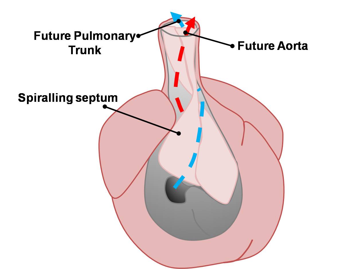
89 KB | category:Heart ILP Truncal and bulbar ridges develop marking the beginning of the division of the outflow tract. | 1 |
| 09:55, 14 March 2010 | Basic Heart Development Timeline.jpg (file) |  |
73 KB | category:Heart ILP | 1 |
| 10:22, 14 March 2010 | Basic Outflow Tract Division.jpg (file) |  |
59 KB | category:Heart ILP Blood flow through the conotruncus is divided to form the pulmonary trunk and aorta. | 1 |
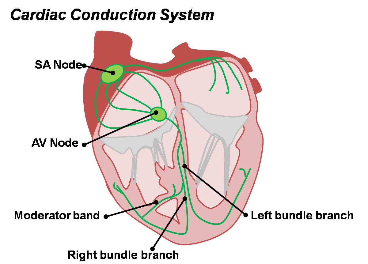
| 12:03, 14 March 2010 | Cardiac Conduction System.jpg (file) |  |
81 KB | category:Heart ILP Cardiac conduction system in the adult heart. | 1 |
| 09:42, 14 March 2010 | Cardiac Embryology ILP Watermark.jpg (file) |  |
78 KB | category:Heart ILP | 1 |
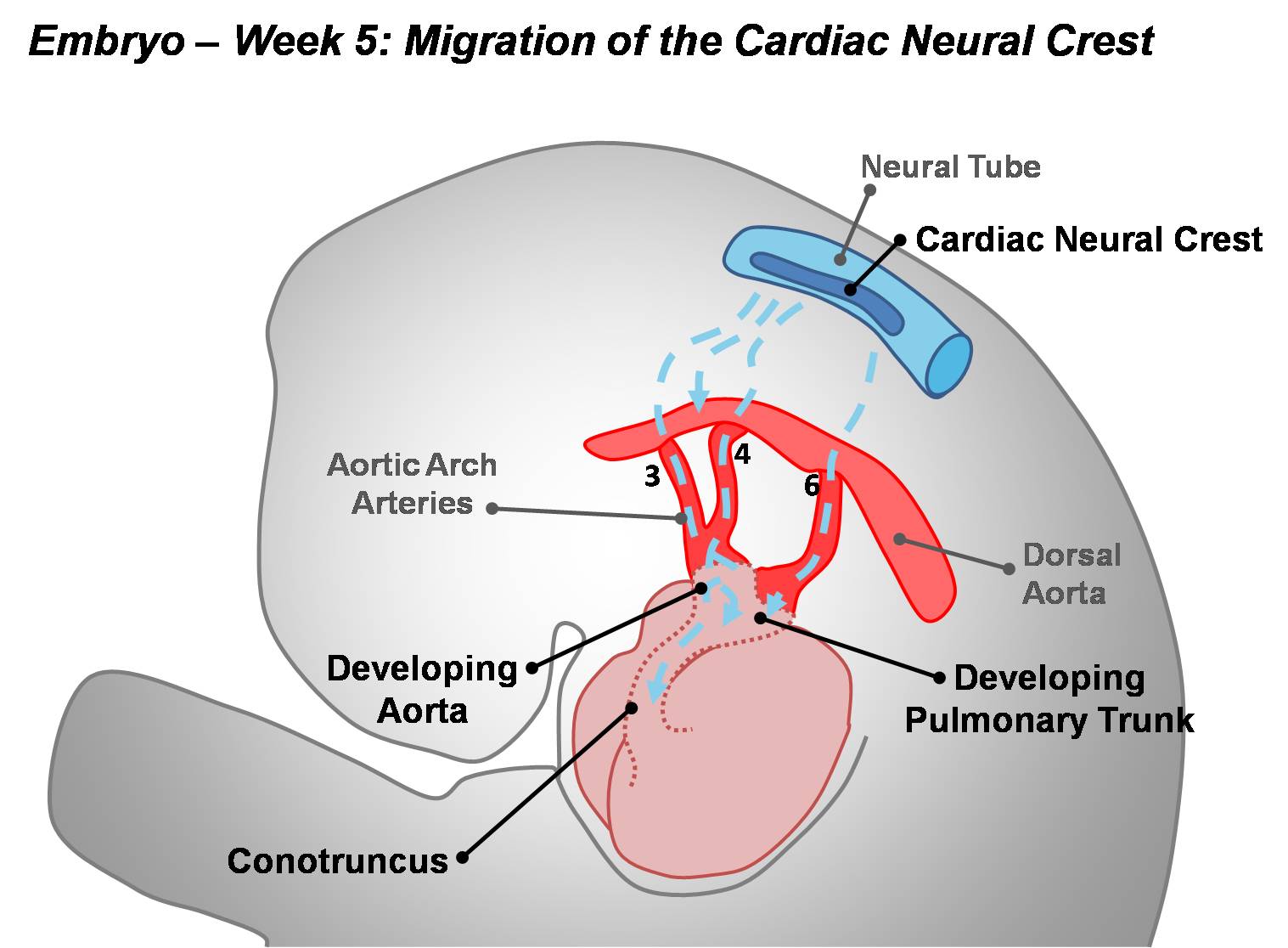
| 11:00, 14 March 2010 | Cardiac Neural Crest Migration.jpg (file) |  |
122 KB | category:Heart ILP In order to complete division of the outflow tract, mesenchyme derived from the cardiac neural crest migrates over the aortic arch arteries to invade the conotruncus. | 1 |
| 11:23, 14 March 2010 | Coarctation of the Aorta.jpg (file) |  |
16 KB | category:Heart ILP | 1 |
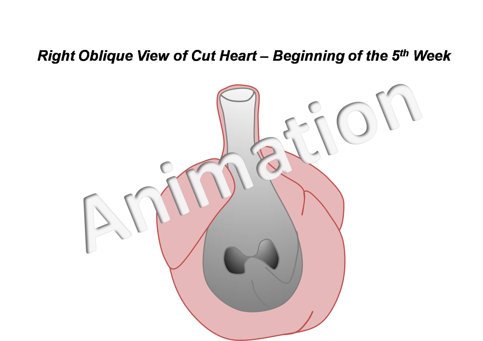
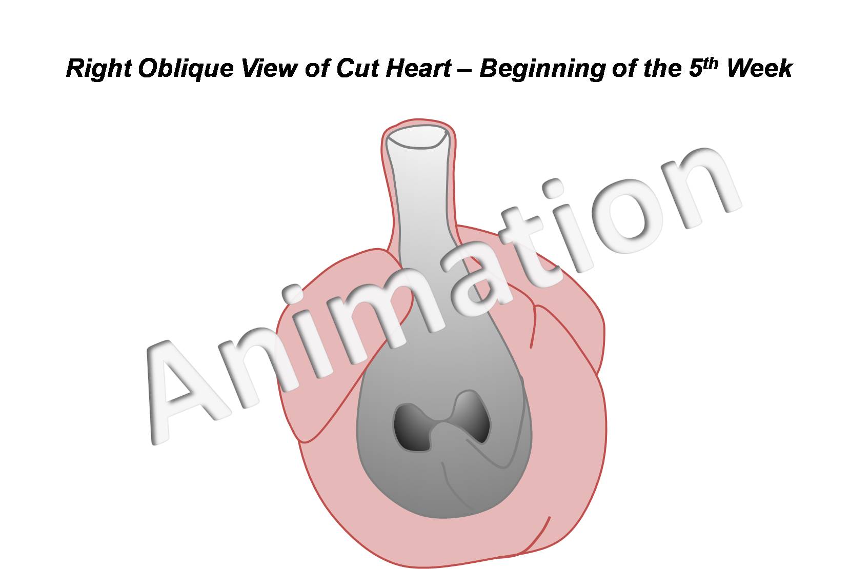
| 10:10, 14 March 2010 | Divisions of Early Heart Tube.jpg (file) |  |
85 KB | {{Template:SEM}} category:Heart ILP | 1 |
| 11:23, 14 March 2010 | Double Outlet Right Ventricle.jpg (file) |  |
16 KB | category:Heart ILP | 1 |
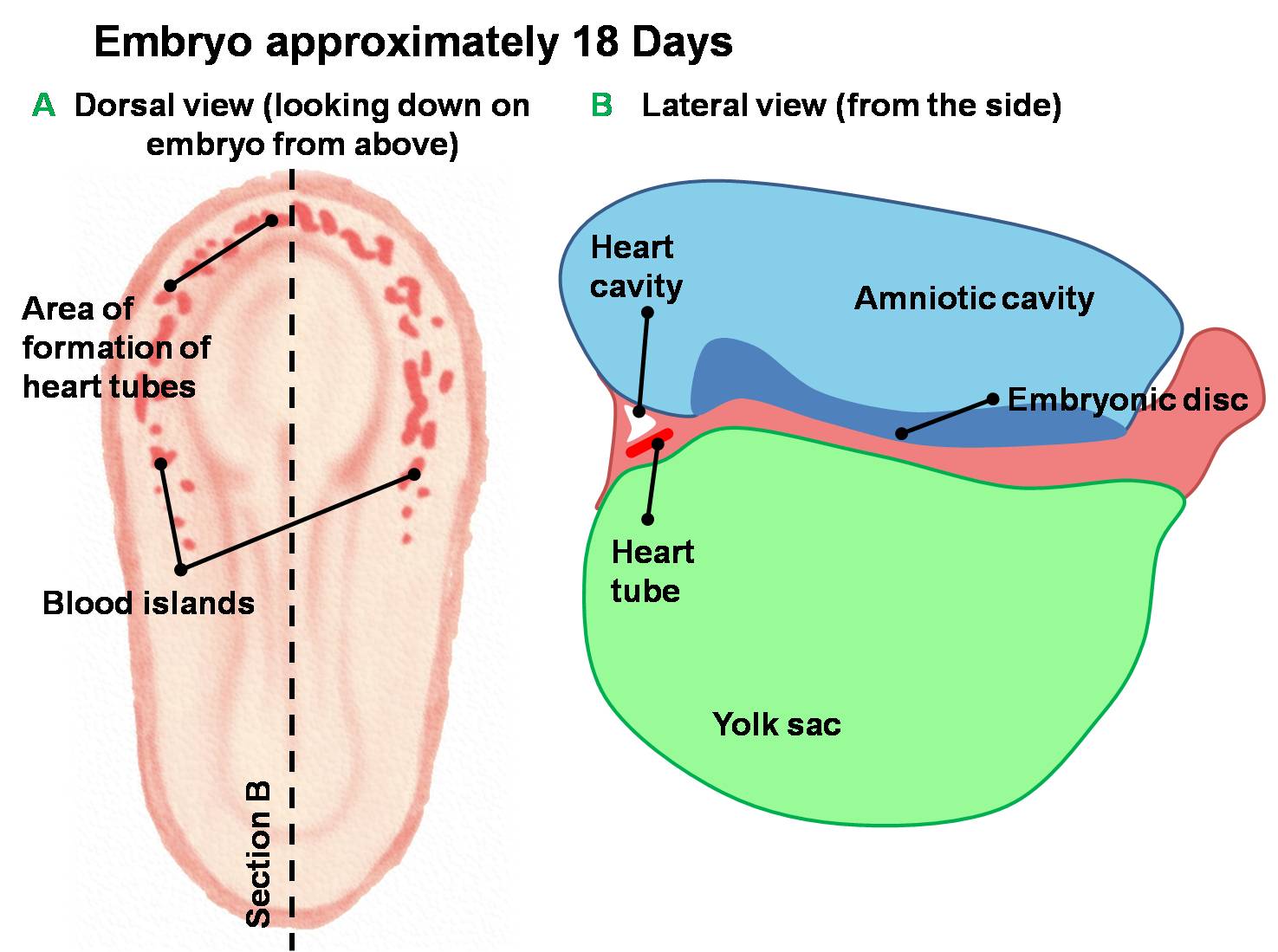
| 10:03, 14 March 2010 | Early Development of Heart Tube.jpg (file) |  |
132 KB | Dorsal and lateral views of the earliest stages of cardiac development in the human embryo. Angiogenesis creates blood islands throughout the embryo during the third week of development. Angioblastic cords form in the cardiogenic mesoderm and canalise to | 1 |
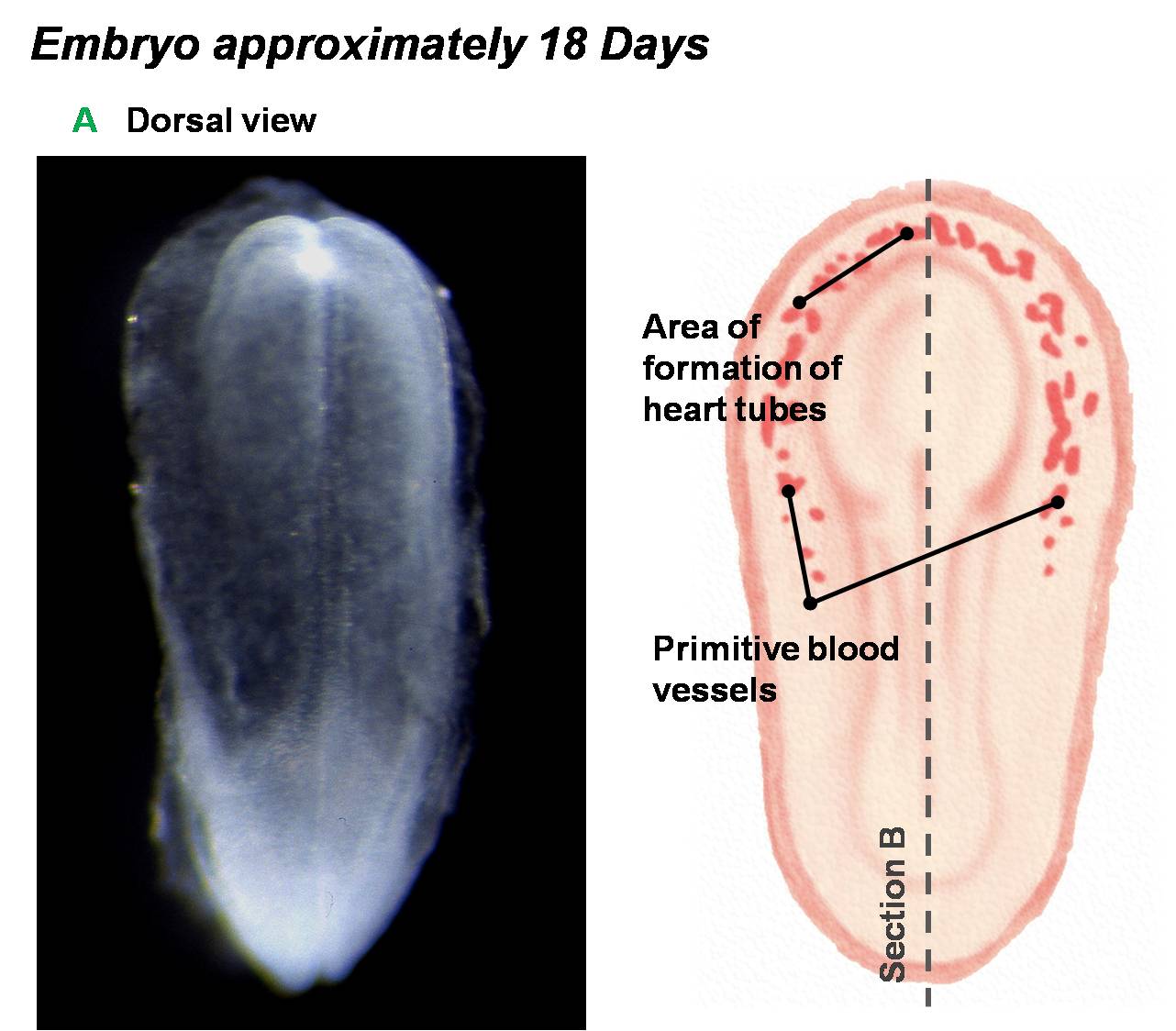
| 10:37, 14 March 2010 | Early Heart Tube (Dorsal).jpg (file) | .jpg) |
111 KB | category:Heart ILP Angiogenesis throughout the embryo allows for the development of angioblastic cords in the cardiogenic mesoderm of the embryo. | 1 |
| 10:38, 14 March 2010 | Early Heart Tube (Lateral).jpg (file) | .jpg) |
110 KB | category:Heart ILP Angiogenesis throughout the embryo allows for the development of angioblastic cords in the cardiogenic mesoderm of the embryo. | 1 |
| 11:39, 14 March 2010 | Embryonic Cardiovascular System (Drawing).jpg (file) | .jpg) |
60 KB | category:Heart ILP | 1 |
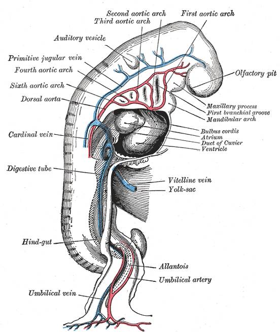
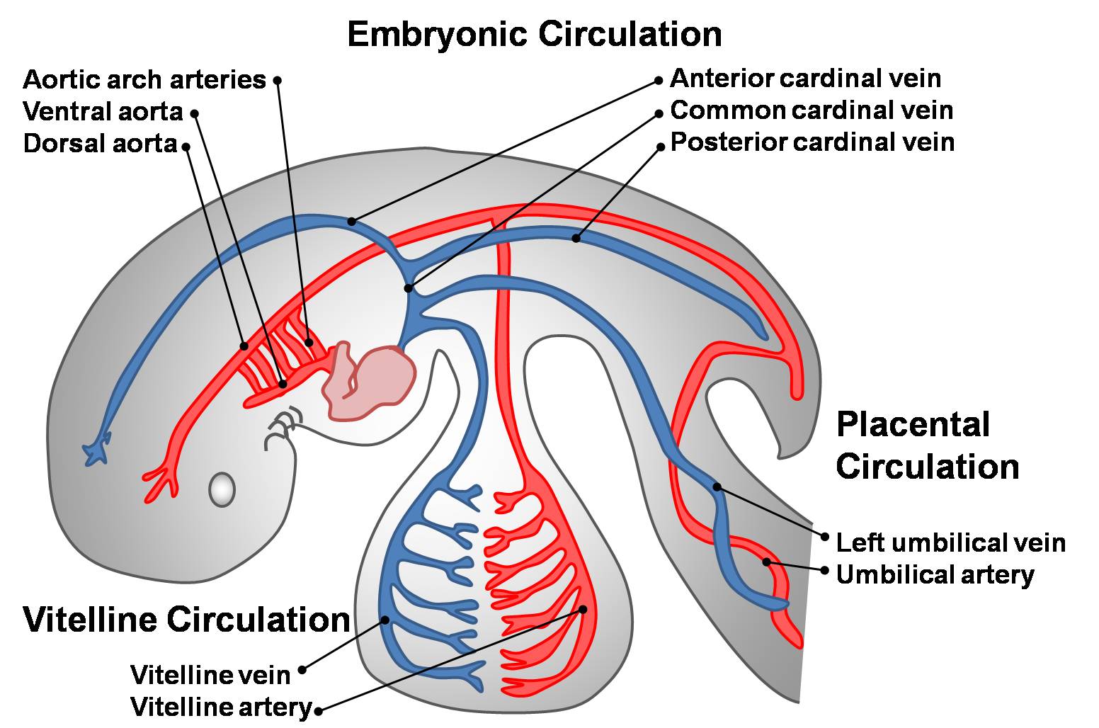
| 10:28, 14 March 2010 | Embryonic Circulations.jpg (file) |  |
171 KB | category:Heart ILP The three early embryonic circulations. Three paired veins drain into the primordial heart tube: vitelline veins (returning poorly oxygenated blood from the yolk sac), umbilical veins (carrying well-oxygenated blood from the primor | 1 |
| 10:56, 14 March 2010 | Embryonic Heart Blood Flow.jpg (file) |  |
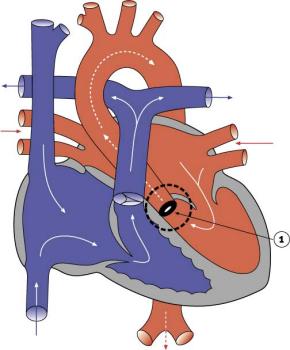
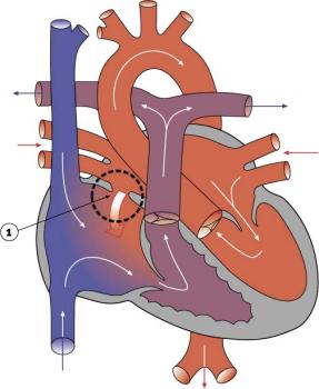
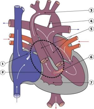
101 KB | category:Heart ILP Oxygenated (from the placenta) and non-oxygenated (from the lower body) blood enters the right atrium via the sinus venosus. Part of this blood travels to the right ventricle and then through the pulmonary circulation. The rest of | 1 |
| 10:29, 14 March 2010 | Fetal Circulation Pathway.jpg (file) |  |
112 KB | category:Heart ILP | 1 |
| 11:24, 14 March 2010 | Functional Hypoplastic Left Heart.jpg (file) |  |
18 KB | category:Heart ILP | 1 |
| 11:48, 8 September 2009 | HeartILP-draft003.jpg (file) |  |
85 KB | Early divisions of the heart tube. category:HeartILP | 1 |
| 11:50, 8 September 2009 | HeartILP-draft004.jpg (file) | 70 KB | Looping of the heart tube. category:HeartILP | 1 | |
| 11:44, 8 September 2009 | HeartILP001.jpg (file) |  |
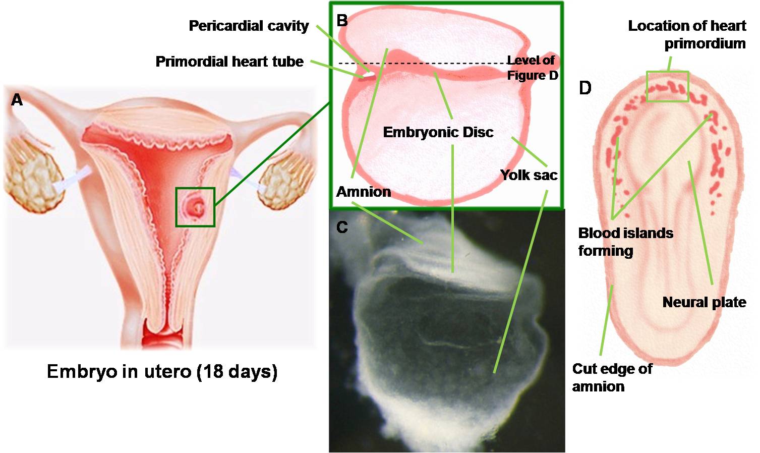
125 KB | Embryo at approximately 18 days showing early angiogenesis and the development of the primordial heart tubes in the cardiogenic region. Brightfield image © Sulik. category:HeartILP | 1 |
| 11:46, 8 September 2009 | HeartILP002.jpg (file) |  |
60 KB | Endocardial tubes fuse together to form the primordial heart tube. category:HeartILP | 1 |
| 08:58, 29 September 2009 | HeartILP draft AVcanalsept.jpg (file) |  |
93 KB | Division of the atrioventricular canal occurs via growth and fusion of the dorsal and ventral (or superior and inferior) endocardial cushions. category:HeartILP | 1 |
| 10:02, 22 September 2009 | HeartILP draft HeartTubeDorsal.jpg (file) |  |
111 KB | Angiogenesis throughout the embryo allows for the development of angioblastic cords in the cardiogenic mesoderm of the embryo. {{Template:SEM}} category:HeartILP | 1 |
| 08:35, 15 October 2009 | HeartILP draft Navigation2.jpg (file) | 52 KB | 1 | ||
| 09:06, 29 September 2009 | HeartILP draft aandvsept.jpg (file) |  |
113 KB | The embryonic heart begins to resemble the adult heart as septation is complete. A right-to-left shunt exists between the right and left atria via the foramen ovale. Both muscular and membranous portions of the ventricular septum are developed, with signi | 1 |
| 11:24, 22 September 2009 | HeartILP draft animpic005.jpg (file) |  |
49 KB | Craniocaudal patterning of cardiogenic progenitors in the epiblast and primitive streak; these migrate through the primitive streak to be organised mediolaterally in the cardiogenic mesoderm. category:HeartILP | 1 |
| 11:32, 22 September 2009 | HeartILP draft animpic006.jpg (file) |  |
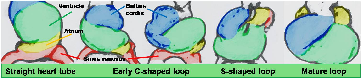
74 KB | Heart looping from ventral, right lateral and left lateral views with the addition of myocardium throughout looping and intrinsic and extrinsic forces controlling looping shown. category:HeartILP | 1 |
| 21:59, 28 September 2009 | HeartILP draft animpic010.jpg (file) |  |
59 KB | Animation of folding of the embryo resulting in fusion of the endocardial heart tubes. category:HeartILP | 1 |
| 22:03, 28 September 2009 | HeartILP draft animpic011.jpg (file) |  |
82 KB | Animation of division of the outflow tract. category:HeartILP | 1 |
| 22:04, 28 September 2009 | HeartILP draft animpic012.jpg (file) |  |
82 KB | Animation of division of the outflow tract. category:HeartILP | 1 |
| 11:06, 5 November 2009 | HeartILP draft atimeline.jpg (file) |  |
158 KB | category:HeartILP | 1 |
| 09:00, 29 September 2009 | HeartILP draft atrialsept1.jpg (file) |  |
90 KB | During septation, the septum primum develops in the roof of the atrium, forming the early left and right atria. The opening of the sinus venosus has shifted to be incorporated into the right atrium. category:HeartILP | 1 |
| 09:03, 29 September 2009 | HeartILP draft atrialsept2.jpg (file) |  |
97 KB | Apoptotic induced perforations appear in the fused septum primum and form the foramen secundum. The septum secundum also begins to grow on the right of the septum primum. The muscular part of the interventricular septum develops concurrently. [[category: | 1 |
| 11:31, 2 October 2009 | HeartILP draft avvalves.jpg (file) |  |
69 KB | Development of the atrioventricular septum over weeks four and five. The right and left atrioventricular canals are remodelled to later become the atrioventricular (tricuspid and mitral) valves. category:HeartILP | 1 |
| 11:12, 5 November 2009 | HeartILP draft btimeline.jpg (file) |  |
73 KB | 2 | |
| 11:09, 29 September 2009 | HeartILP draft coarctationoftheaorta.jpg (file) |  |
16 KB | category:HeartILP | 1 |
| 11:11, 29 September 2009 | HeartILP draft funchlh.jpg (file) |  |
18 KB | category:HeartILP | 1 |
| 11:00, 29 September 2009 | HeartILP draft grayembryoniccirc.jpg (file) |  |
60 KB | category:HeartILP | 1 |
| 10:15, 22 September 2009 | HeartILP draft hearttubesegments.jpg (file) |  |
63 KB | As the tubular heart grows it develops dilations and constrictions which form the truncus arteriosus, bulbus cordis, primitive ventricle, primitive atrium and sinus venosus. {{Template:SEM}} category:HeartILP | 1 |