Uploads by S8600021
From Embryology
This special page shows all uploaded files.
| Date | Name | Thumbnail | Size | Description | Versions |
|---|---|---|---|---|---|
| 22:26, 13 February 2012 | Keibel Mall table01.jpg (file) |  |
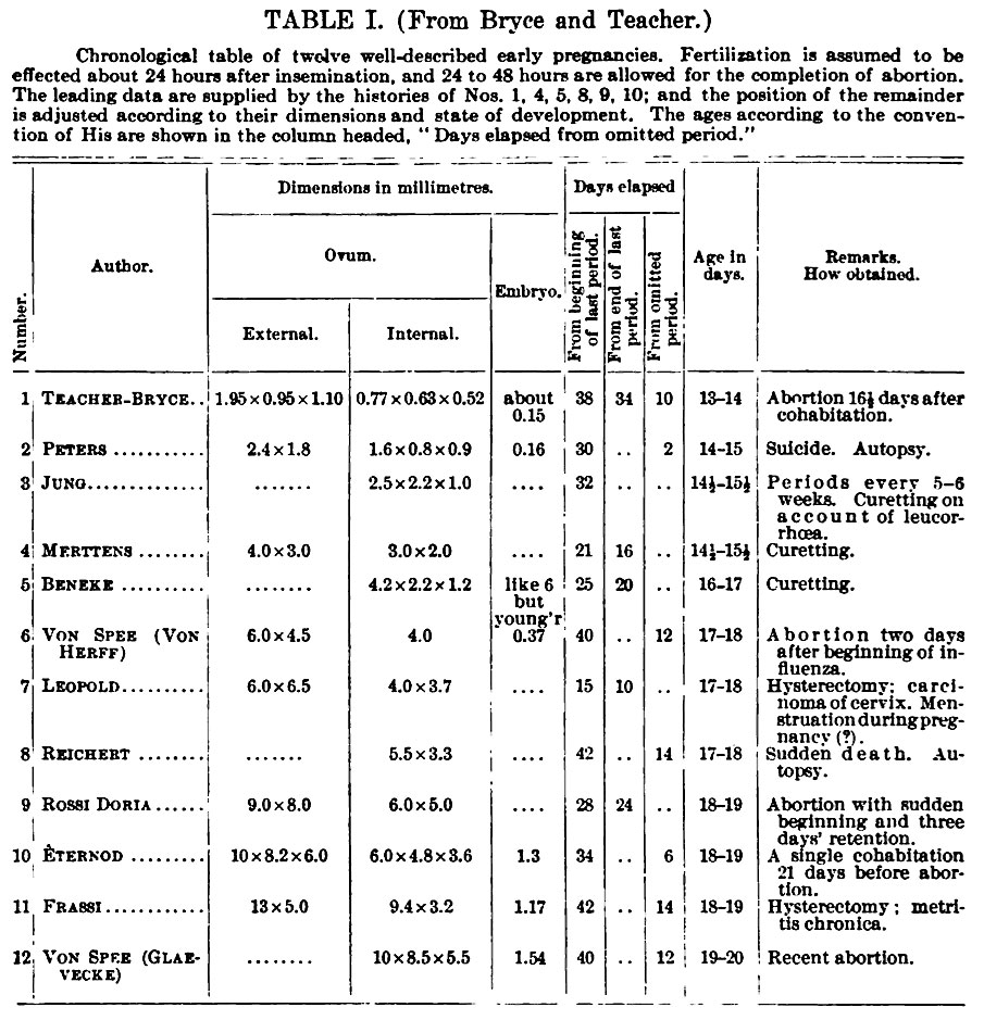
225 KB | ==Table I. Chronological Table of Twelve Well-Described Early Pregnancies== From Bryce and Teacher. Fertilization is assumed to be affected about 24 hours after insemination, and 24 to 48 hours are allowed for the completion of abortion. Keibel_Mall_ta | 1 |
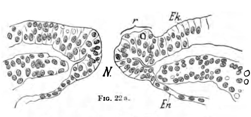
| 22:23, 13 February 2012 | Keibel Mall 022-026.jpg (file) |  |
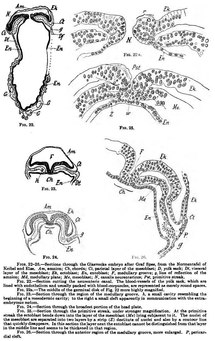
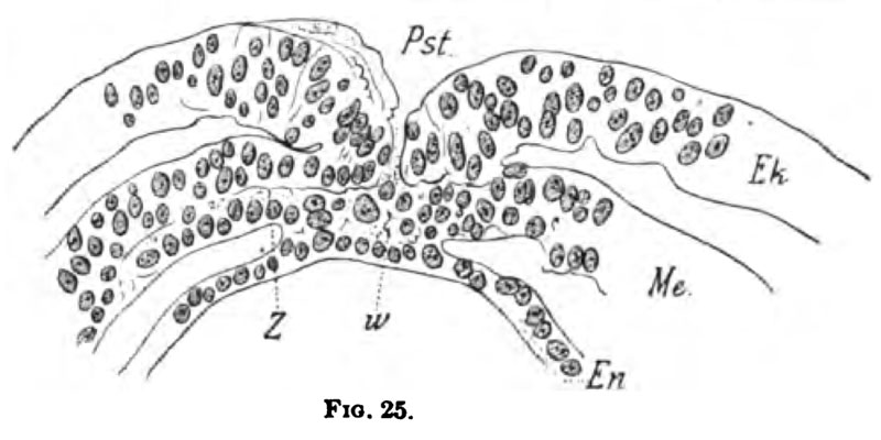
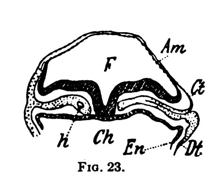
226 KB | ==Fig. 22. to Fig. 26. Section of the Embryo== Keibel_Mall_022.jpg {{Keibel_Mall Images}} Category:Human | 1 |
| 22:22, 13 February 2012 | Keibel Mall 025.jpg (file) |  |
56 KB | ==Fig. 25. Section of the Embryo== Keibel_Mall_022.jpg {{Keibel_Mall Images}} Category:Human | 1 |
| 22:21, 13 February 2012 | Keibel Mall 024.jpg (file) |  |
28 KB | ==Fig. 22. Section of the Embryo== Keibel_Mall_022.jpg {{Keibel_Mall Images}} Category:Human | 1 |
| 22:21, 13 February 2012 | Keibel Mall 023.jpg (file) |  |
34 KB | ==Fig. 22. Section of the Embryo== Keibel_Mall_022.jpg {{Keibel_Mall Images}} Category:Human | 1 |
| 22:20, 13 February 2012 | Keibel Mall 022a.jpg (file) |  |
52 KB | ==Fig. 22. Section of the Embryo== Keibel_Mall_022.jpg {{Keibel_Mall Images}} Category:Human | 1 |
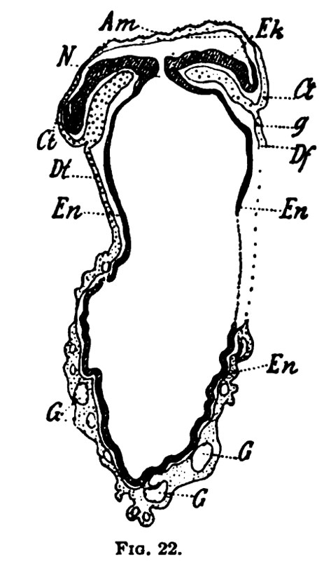
| 22:20, 13 February 2012 | Keibel Mall 022.jpg (file) |  |
63 KB | ==Fig. 22. Section of the Embryo== Keibel_Mall_022.jpg {{Keibel_Mall Images}} Category:Human | 1 |
| 22:19, 13 February 2012 | Keibel Mall 021.jpg (file) |  |
113 KB | ==Fig. 21. Three Views of the Glaevecke Embryo of Graf Spee== {{Keibel_Mall Images}} Category:Human | 1 |
| 22:14, 13 February 2012 | Keibel Mall 020.jpg (file) |  |
80 KB | ==Fig. 20. Median Sagittal Section of the Glaevecke Embryo of Graf Spee== {{Keibel_Mall Images}} Category:Human | 1 |
| 22:11, 13 February 2012 | Keibel Mall 019.jpg (file) |  |
40 KB | ==Fig. 19. The Glaevecke Embryo of Graf Spee== Seen from the dorsal surface. From a model. {{Keibel_Mall Images}} Category:Human | 1 |
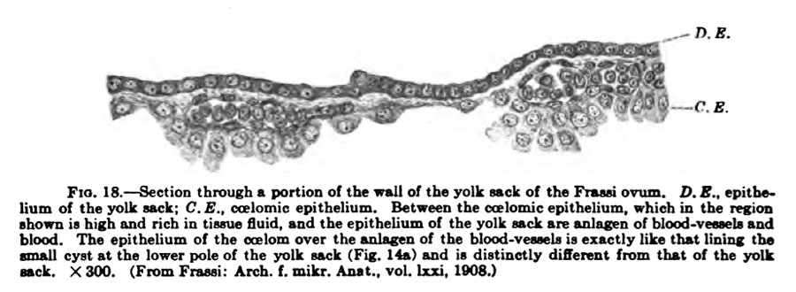
| 22:10, 13 February 2012 | Keibel Mall 018.jpg (file) |  |
70 KB | ==Fig. 18. Section of the Yolk Sack of the Frassi Ovum== {{Keibel_Mall Images}} Category:Human | 1 |
| 22:09, 13 February 2012 | Keibel Mall 017.jpg (file) |  |
74 KB | ==Fig. 17. Section of the Frassi Ovum== {{Keibel_Mall Images}} Category:Human | 1 |
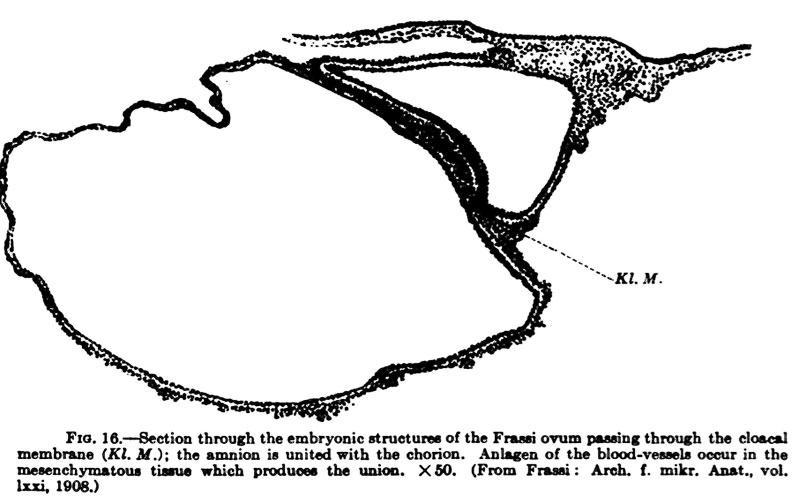
| 22:08, 13 February 2012 | Keibel Mall 016.jpg (file) |  |
67 KB | ==Fig. 16. Section of the Frassi Ovum== {{Keibel_Mall Images}} Category:Human | 1 |
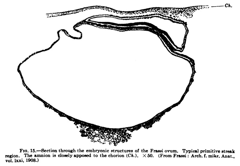
| 22:07, 13 February 2012 | Keibel Mall 015.jpg (file) |  |
62 KB | ==Fig. 15. Section of the Frassi Ovum== {{Keibel_Mall Images}} Category:Human | 1 |
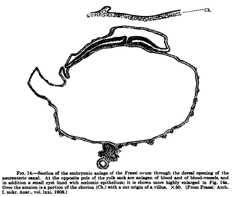
| 21:24, 13 February 2012 | Keibel Mall 014.jpg (file) |  |
80 KB | ==Fig. 14. Section of the Frassi Ovum== {{Keibel_Mall Images}} Category:Human | 1 |
| 21:24, 13 February 2012 | Keibel Mall 014a.jpg (file) |  |
70 KB | ==Fig. 14a. Section of the Frassi Ovum== {{Keibel_Mall Images}} Category:Human | 1 |
| 21:21, 13 February 2012 | Keibel Mall 013.jpg (file) |  |
60 KB | ==Fig. 13. Section of the Frassi Ovum== {{Keibel_Mall Images}} Category:Human | 1 |
| 21:20, 13 February 2012 | Keibel Mall 012.jpg (file) |  |
95 KB | ==Fig. 10. Figure of the Embryonic Shield of the Frassi Ovum== {{Keibel_Mall Images}} Category:Human | 1 |
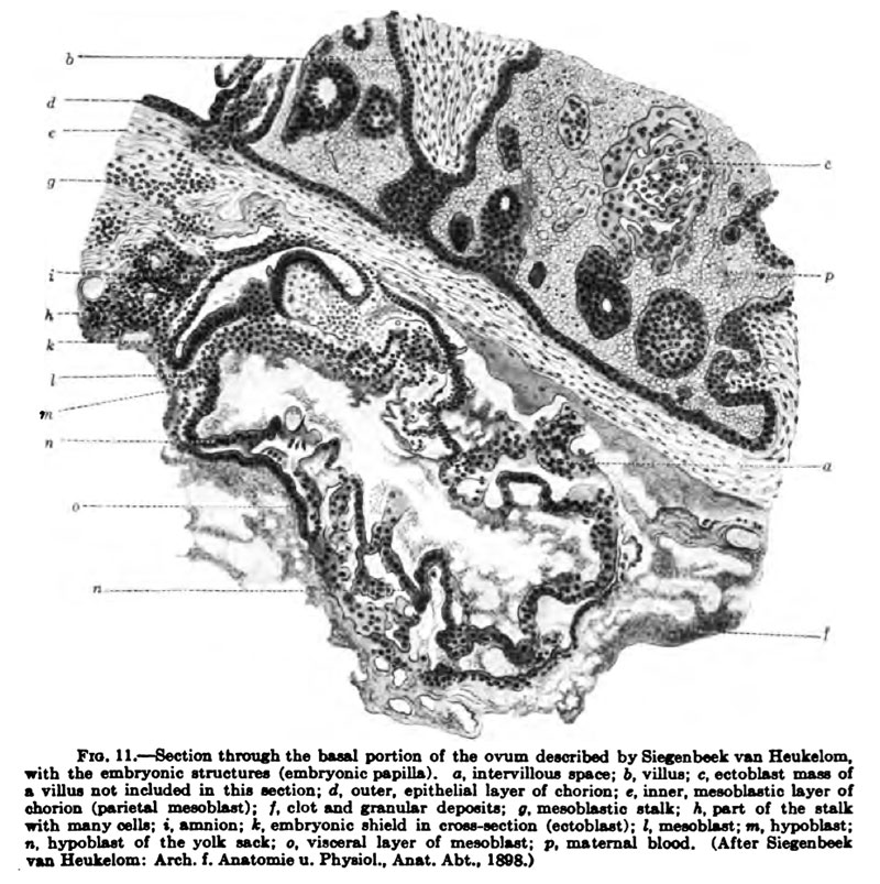
| 21:19, 13 February 2012 | Keibel Mall 011.jpg (file) |  |
171 KB | ==Fig. 11. Section through the Basal Portion of the Ovum== Described by Siegenbeck van Heukelom. {{Keibel_Mall Images}} Category:Human | 1 |
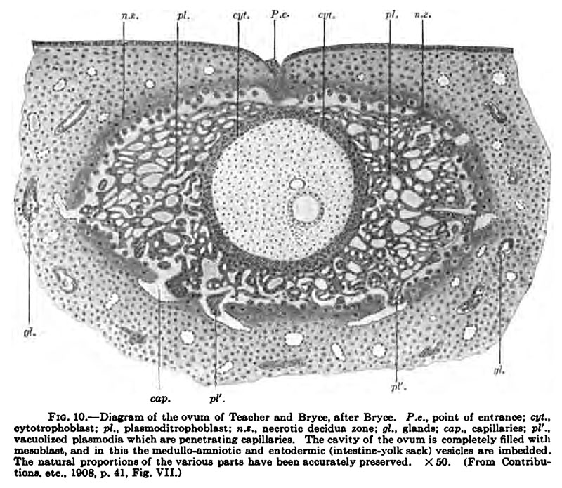
| 19:43, 13 February 2012 | Keibel Mall 010.jpg (file) |  |
154 KB | ==Fig. 10. Diagram of the Ovum of Teacher and Bryce== Keibel Mall 010.jpg {{Keibel_Mall Images}} Category:Human | 1 |
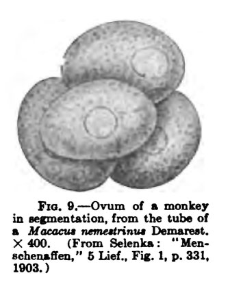
| 19:36, 13 February 2012 | Keibel Mall 009.jpg (file) |  |
27 KB | ==Fig. 9. Ovum from a Monkey== Keibel Mall 001.jpg {{Keibel_Mall Images}} Category:Monkey | 1 |
| 20:12, 12 February 2012 | Keibel Mall 001.jpg (file) |  |
138 KB | ==Fig. 1. == {{Keibel_Mall Images}} | 1 |
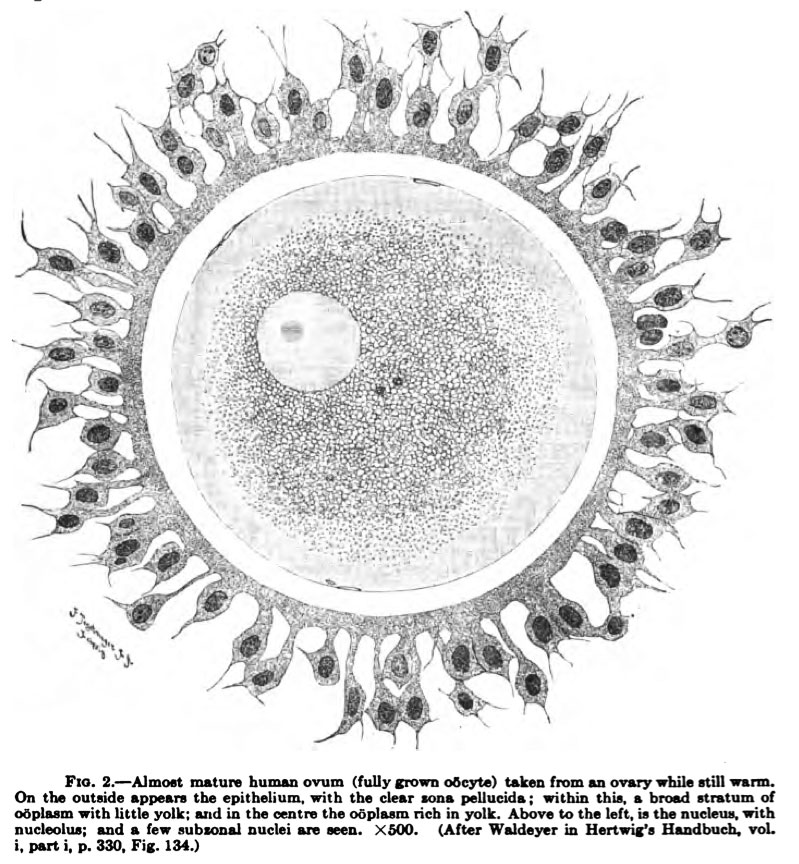
| 20:11, 12 February 2012 | Keibel Mall 002.jpg (file) |  |
167 KB | ==Fig. 2. == {{Keibel_Mall Images}} | 1 |
| 20:11, 12 February 2012 | Keibel Mall 003.jpg (file) |  |
26 KB | ==Fig. 3. == {{Keibel_Mall Images}} | 1 |
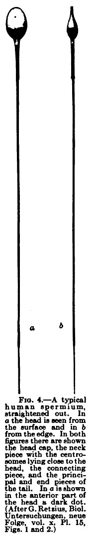
| 20:11, 12 February 2012 | Keibel Mall 004.jpg (file) |  |
42 KB | ==Fig. 4. == {{Keibel_Mall Images}} | 1 |
| 20:11, 12 February 2012 | Keibel Mall 005.jpg (file) |  |
143 KB | ==Fig. 5. == {{Keibel_Mall Images}} | 1 |
| 20:10, 12 February 2012 | Keibel Mall 006.jpg (file) |  |
77 KB | ==Fig. 6. == {{Keibel_Mall Images}} | 1 |
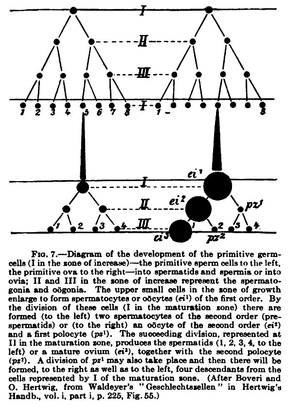
| 20:10, 12 February 2012 | Keibel Mall 007.jpg (file) |  |
152 KB | ==Fig. 7. == {{Keibel_Mall Images}} | 1 |
| 18:04, 12 February 2012 | Keibel Mall 008.jpg (file) |  |
161 KB | ==Fig. 8. Diagram showing a comparison of the testis and the ovary== Diagram showing a comparison of the testis and the ovary (based on the results of Winiwarter and Waldeyer). The germinal epithelium of the texts corresponds to 1-3 of the ovary. {{Keib | 1 |
| 17:00, 12 February 2012 | Mouse in vitro follicle 06.jpg (file) |  |
120 KB | ==In Vitro Grown Mouse Non-Antral Follicle== Appearance of in vitro grown mouse follicles a, c, e, ZP: zona pellucida; b: basement membrane. Mouse in vitro follicle 01.jpg ===Reference=== <pubmed>21232101</pubmed>| [http://www.ncbi.nlm.nih.gov/pmc/artic | 1 |
| 16:57, 12 February 2012 | Mouse in vitro follicle 05.jpg (file) |  |
82 KB | ==In Vitro Grown Mouse Antral Follicle== Appearance of in vitro grown mouse follicles a, c, e, ZP: zona pellucida; b: basement membrane. Mouse in vitro follicle 01.jpg ===Reference=== <pubmed>21232101</pubmed>| [http://www.ncbi.nlm.nih.gov/pmc/articles/ | 1 |
| 16:56, 12 February 2012 | Mouse in vitro follicle 04.jpg (file) |  |
81 KB | ==In Vitro Grown Mouse Follicle Electron Micrograph== Appearance of in vitro grown mouse follicles a, c, e, ZP: zona pellucida; b: basement membrane. Mouse in vitro follicle 01.jpg ===Reference=== <pubmed>21232101</pubmed>| [http://www.ncbi.nlm.nih.gov/ | 1 |
| 16:55, 12 February 2012 | Mouse in vitro follicle 03.jpg (file) |  |
99 KB | ==In Vitro Grown Mouse Non-Antral Follicle== Appearance of in vitro grown mouse follicles a, c, e, ZP: zona pellucida; b: basement membrane. Mouse in vitro follicle 01.jpg ===Reference=== <pubmed>21232101</pubmed>| [http://www.ncbi.nlm.nih.gov/pmc/artic | 1 |
| 16:54, 12 February 2012 | Mouse in vitro follicle 02.jpg (file) |  |
120 KB | ==In Vitro Grown Mouse Antral Follicle== Appearance of in vitro grown mouse follicles a, c, e, ZP: zona pellucida; b: basement membrane. Mouse in vitro follicle 01.jpg ===Reference=== <pubmed>21232101</pubmed>| [http://www.ncbi.nlm.nih.gov/pmc/articles/ | 1 |
| 16:50, 12 February 2012 | Mouse in vitro follicle 01.jpg (file) |  |
72 KB | ==In Vitro Grown Mouse Follicle== Appearance of in vitro grown mouse follicles a, c, e, ZP: zona pellucida; b: basement membrane. Mouse in vitro follicle 01.jpg ===Reference=== [http://www.ncbi.nlm.nih.gov/pmc/articles/PMC3033320 PMC3033320] This is | 1 |
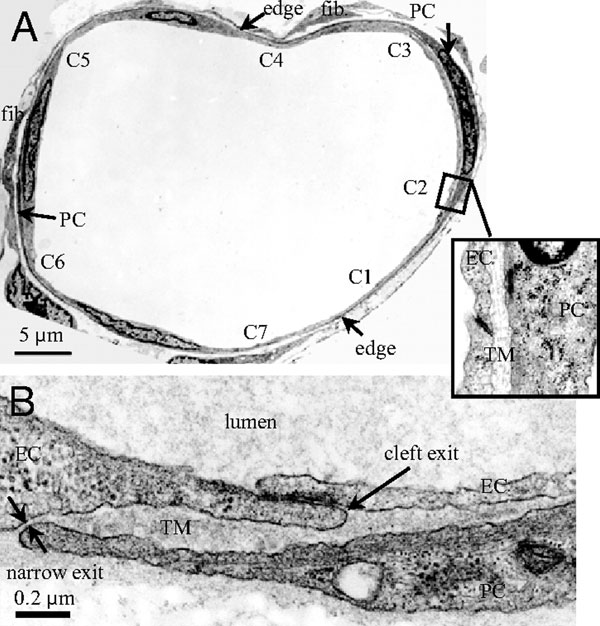
| 13:40, 12 February 2012 | Venule microvessel EM.jpg (file) |  |
91 KB | ==Electron microscope images of a rat venular microvessel== (A) Cross-section of a venular microvessel (VM) in rat mesentery. There are seven endothelial cell (EC) clefts, C1-C7, with all of the cleft exits covered by neighboring pericytes (PCs). PCs are | 1 |
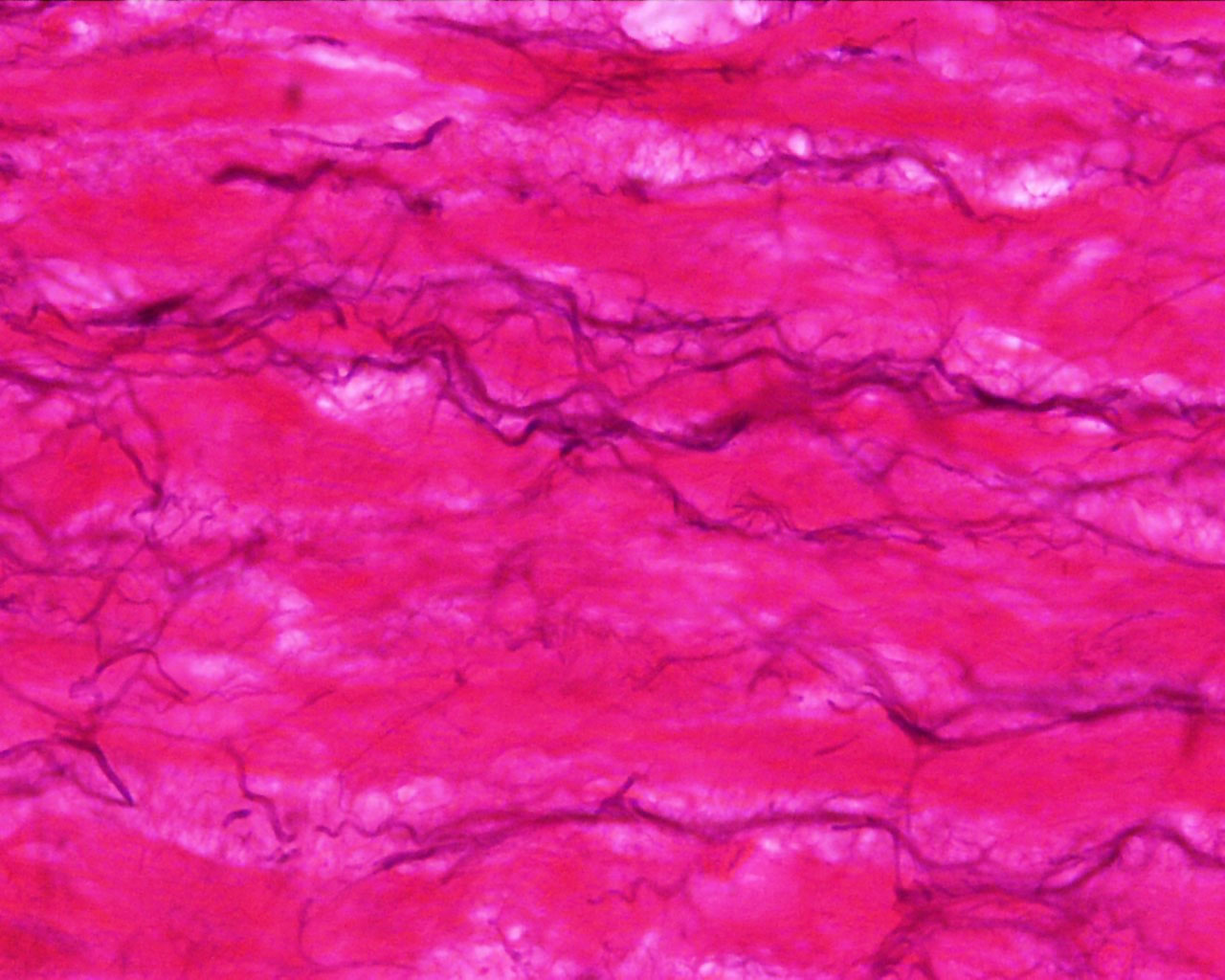
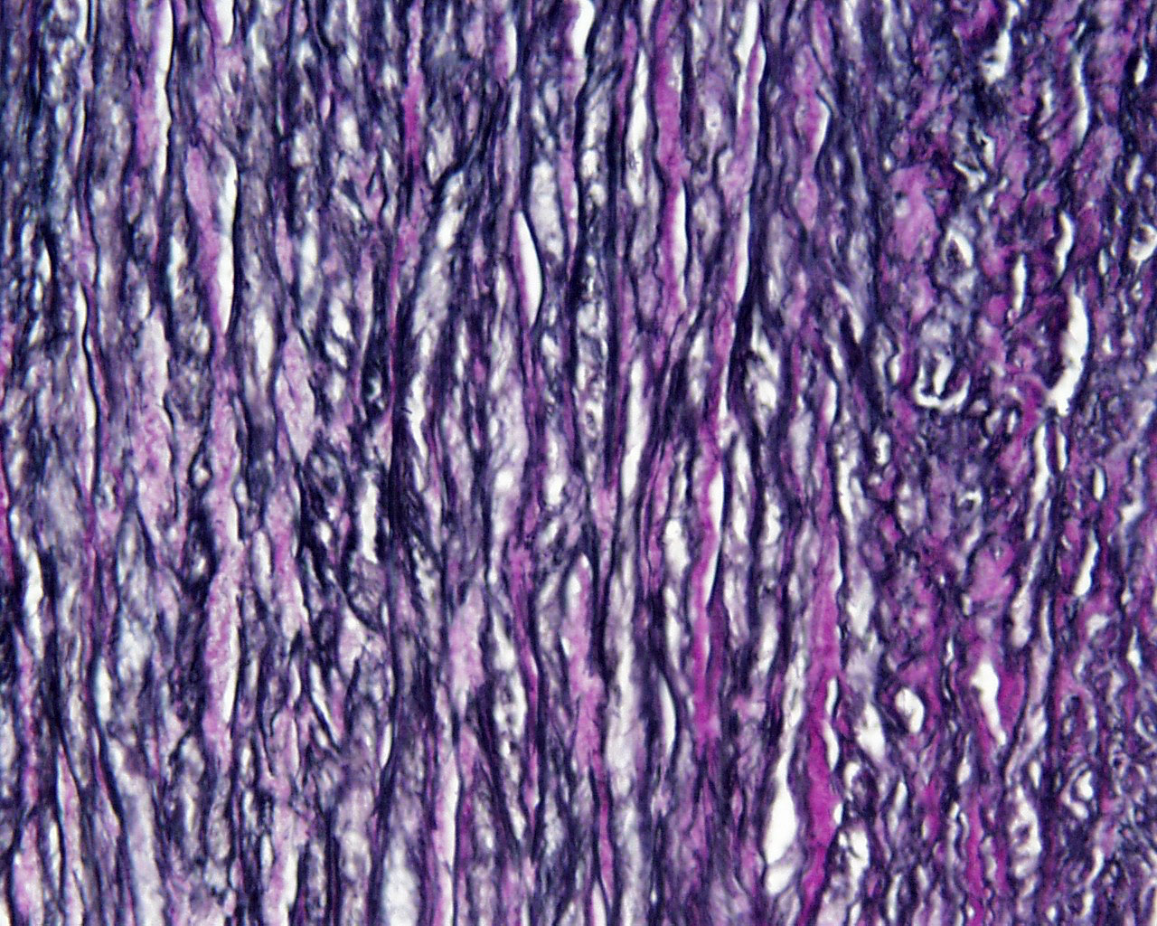
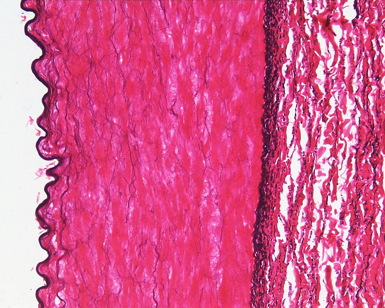
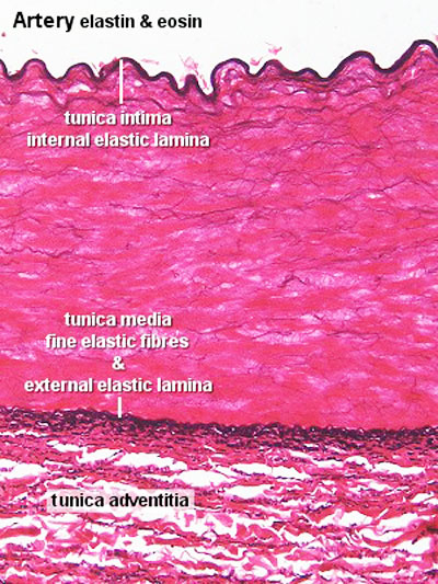
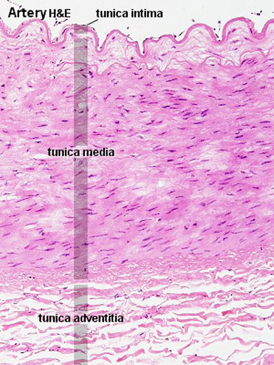
| 13:06, 12 February 2012 | Artery histology 12.jpg (file) |  |
206 KB | ==Artery Histology== * muscular artery, tunica media, elastic fibres. Vessels close to the heart (aorta, pulmonary trunk and the larger arteries that originate from them) are elastic arteries, the tunica intima of elastic arteries will be thicker than in | 1 |
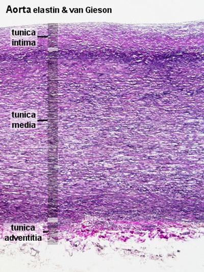
| 12:58, 12 February 2012 | Artery histology 16.jpg (file) |  |
409 KB | ==Aorta Histology== Vessels close to the heart (aorta, pulmonary trunk and the larger arteries that originate from them) are elastic arteries, the tunica intima of elastic arteries will be thicker than in other arteries. Elastin stain will show: * inner | 1 |
| 10:34, 12 February 2012 | Stage23 bf8.jpg (file) |  |
43 KB | == Human Embryo Carnegie Stage 23== Carnegie Stage 23 (day 56) Carnegie Collection Embryo No.4570 Left side view Facts: Week 8, 56 - 60 days, 27 - 31 mm Features: scalp vascular plexus, eylid, eye, nose, auricle of external ear, mouth, sholder, arm, | 1 |
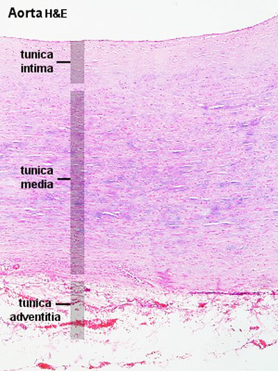
| 10:30, 12 February 2012 | Artery histology 15.jpg (file) |  |
343 KB | ==Aorta Histology== * aorta, human HE * elastic artery, tunica media, elastic fibres {{Blood Vessel Histology}} {{Blue Histology}} | 1 |
| 10:23, 12 February 2012 | Artery histology 14.jpg (file) |  |
466 KB | ==Artery Histology== Vessels close to the heart (aorta, pulmonary trunk and the larger arteries that originate from them) are elastic arteries, the tunica intima of elastic arteries will be thicker than in other arteries. Elastin stain will show: * inn | 1 |
| 10:19, 12 February 2012 | Artery histology 13.jpg (file) |  |
474 KB | ==Artery Histology== Vessels close to the heart (aorta, pulmonary trunk and the larger arteries that originate from them) are elastic arteries, the tunica intima of elastic arteries will be thicker than in other arteries. Elastin stain will show: * inner | 1 |
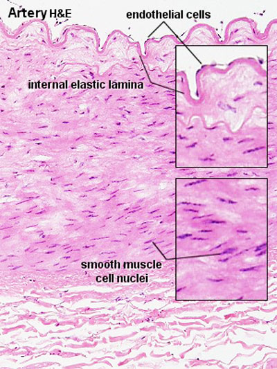
| 10:15, 12 February 2012 | Artery histology 11.jpg (file) |  |
344 KB | ==Artery Histology== {{Blood Vessel Histology}} {{Blue Histology}} Aty10he.jpg Artery histology 01.jpg | 1 |
| 08:22, 12 February 2012 | Artery histology 06.jpg (file) |  |
91 KB | ==Aorta Histology== Vessels close to the heart (aorta, pulmonary trunk and the larger arteries that originate from them) are elastic arteries, the tunica intima of elastic arteries will be thicker than in other arteries. Elastin stain will show: * inner | 1 |
| 08:21, 12 February 2012 | Artery histology 05.jpg (file) |  |
72 KB | ==Aorta Histology== {{Blood Vessel Histology}} {{Blue Histology}} | 1 |
| 08:05, 12 February 2012 | Artery histology 03.jpg (file) |  |
89 KB | ==Artery Histology== Elastin stain will show: * inner elastic laminae * outer elastic laminae * fine elastic fibres in the tunica media * coarse elastic fibres between the collagen fibres of the tunica adventitia {{Blood Vessel Histology}} {{Blue His | 1 |
| 07:59, 12 February 2012 | Artery histology 02.jpg (file) |  |
78 KB | ==Artery Histology== {{Blood Vessel Histology}} {{Blue Histology}} Aty11he.jpg | 1 |
| 07:57, 12 February 2012 | Artery histology 01.jpg (file) |  |
80 KB | ==Artery Histology== {{Blood Vessel Histology}} {{Blue Histology}} Aty10he.jpg Artery histology 01.jpg | 1 |
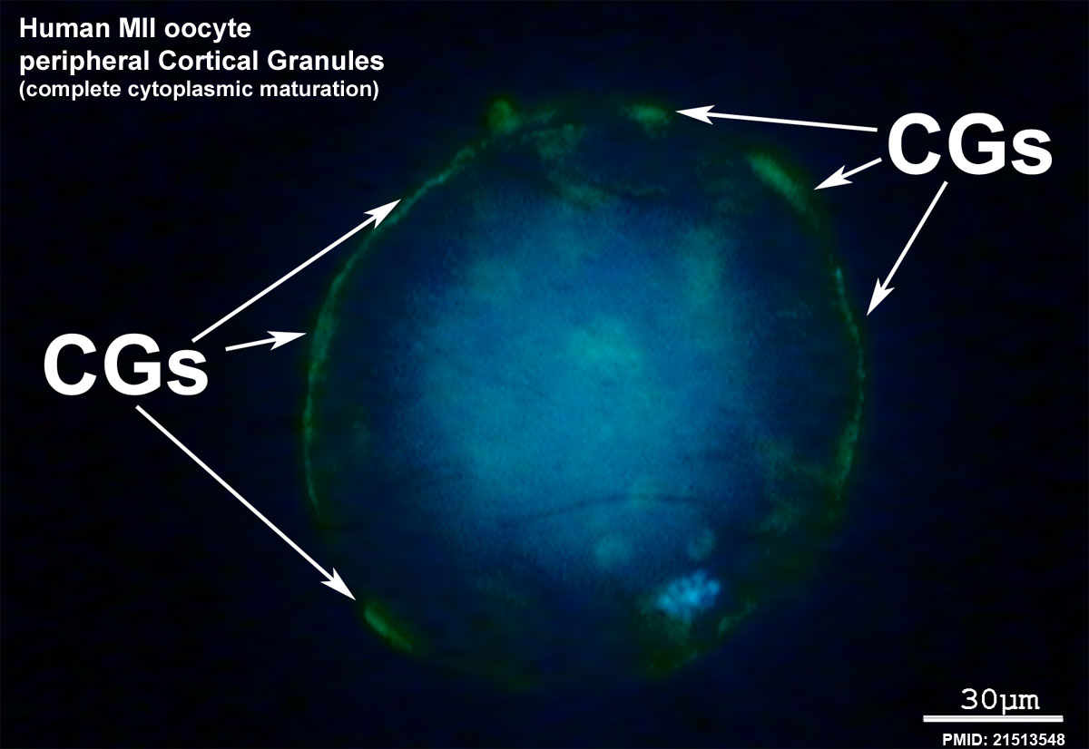
| 15:08, 5 February 2012 | Human MII oocyte 02.jpg (file) |  |
98 KB | ==Human MII Oocyte Cytoplasmic Maturation== Complete cytoplasmic maturation, cortical granules (CGs, green) are located on the MII oocyte periphery. Category:Human Category:Oocyte Category:Fluorescent | 1 |
| 15:06, 5 February 2012 | Human MII oocyte 01.jpg (file) |  |
92 KB | ==Human MII Oocyte Cytoplasmic Maturation== Incomplete cytoplasmic maturation, CGs are both clustered in the center and on the periphery of the MII oocyte. CGs = cortical granules Category:Human Category:Oocyte Category:Fluorescent | 1 |