Uploads by Z3465141
From Embryology
This special page shows all uploaded files.
| Date | Name | Thumbnail | Size | Description | Versions |
|---|---|---|---|---|---|
| 09:24, 24 October 2014 | Horseshoe Kidney.jpg (file) |  |
205 KB | ==Horseshoe Kidney diagnosis by intravenous pyelography== Diagnosis of horseshoe kidney was made by intravenous pyelography. Coronal volume rendering multidetector CT image shows various blood supply to the horseshoe kidney. Right and left renal arter... | 1 |
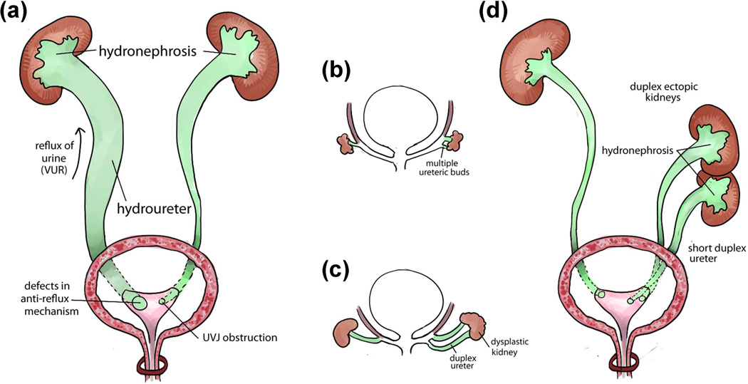
| 10:55, 22 October 2014 | Duplicated ureter .jpg (file) |  |
159 KB | ==Duplicated Ureter== (a) Two main causes of hydronephrosis and hydroureter: vesicoureteral reflux (VUR) caused by defects of anti-reflux mechanism is on the left side and urinary obstruction caused by abnormal structure of the ureterovesical junction... | 1 |
| 10:18, 22 October 2014 | Ureter.jpg (file) |  |
226 KB | ==Urinary tract development and structure== (a) Early development of the urinary tract (fourth week of gestation in humans and E10.5 in mice). An epithelial diverticulum called the ureteric bud (UB) emanates from the Wolffian duct and grows into an ad... | 1 |
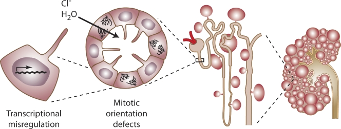
| 07:54, 6 October 2014 | PKD.jpg (file) |  |
163 KB | ==Cyst formation at the level of the cell, nephron, and kidney== Defects in the genes encoding PC1 or PC2 lead to aberrant gene transcription, cell proliferation, and ion secretion, which in turn result in the formation of fluid-filled cysts. As cysts... | 1 |
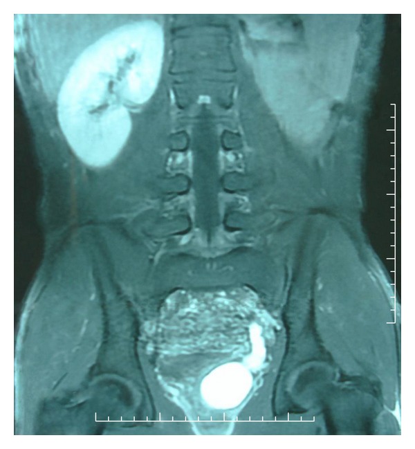
| 14:30, 17 September 2014 | MRI renal agenesis .jpg (file) |  |
67 KB | =Renal agenesis= MRI imaging confirming renal agenesis ==Reference== <pubmed>22606606</pubmed> [http://www.ncbi.nlm.nih.gov/pubmed/22606606] ==Copyright== Copyright © 2011 Youness Ahallal et al. This is an open access article distributed under the... | 2 |
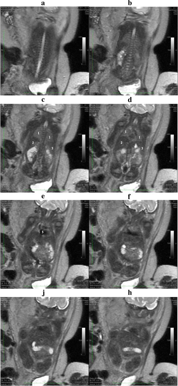
| 10:20, 20 August 2014 | MRI confirming renal agenesis.jpg (file) |  |
251 KB | The image above shows the confirmation of renal agenesis following an MRI. Image was taken of at the 28th week of gestation from a coronal frame from the dorsal to ventral side of the fetus showing from a-h. Image key is as follows: 1 - lung 2 - live... | 1 |