Uploads by Z3418702
From Embryology
This special page shows all uploaded files.
| Date | Name | Thumbnail | Size | Description | Versions |
|---|---|---|---|---|---|
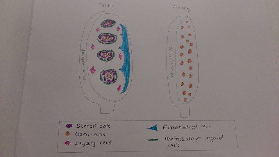
| 13:55, 24 October 2014 | Testis and ovary.jpg (file) |  |
37 KB | ==Ovary and testis cell types== Image indicating the different component cells of the mammalian testis and ovary in gestation. This ia a student drawn representation, adapted from <ref>Dagmar Wilhelm, Jennifer X Yang, Paul Thomas Chapter Three – Mamm... | 1 |
| 08:33, 24 October 2014 | Fetal ovary morphogenesis (mouse).jpg (file) | .jpg) |
93 KB | ==Fetal mouse ovary morphogenesis== Image indicating the different component cells in ovary organogenesis in the fetal mouse. PGCs coalesce with somatic cells derived from (1), (2) or (3) in the diagram. (A)- Ovigerous cord formation by PGCs and somat... | 1 |
| 19:41, 23 October 2014 | Placenta accreta.jpg (file) |  |
98 KB | ==Placenta accreta== This image shows an area of profuse bleeding in the human placenta, with an arrow indicating an area of placenta accreta ===Reference=== <pubmed>21867547</pubmed> '''Copyright''' ©2011 Tikkanen et al; licensee BioMed Central L... | 1 |
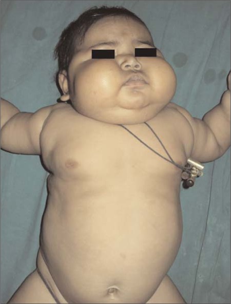
| 18:08, 23 October 2014 | Cushing's syndrome.jpg (file) |  |
62 KB | ==Clinical manifestations of Cushing’s Syndrome== This image shows a child at 3 months of age showing central obesity, moon facies and chubby cheeks; classic signs of Cushing’s Syndrome in infants. ===Reference=== <pubmed>22985617</pubmed> '''Co... | 1 |
| 16:38, 22 October 2014 | Placenta schematic.jpg (file) |  |
121 KB | ==Diagramatic representation of the human placenta== This image indicates the different components of the mature placenta; fetal placenta, decidua, chorionic villous, intervillous space and maternal spiral arteries that supply the fetus. ===Reference... | 1 |
| 10:26, 22 October 2014 | Fetal adrenal gland.png (file) |  |
7.7 MB | ==The fetal adrenal gland== A and B- Fetal adrenal gland of gestational weeks 24 and 36 shown by a transverse MRI C and E- Show the right adrenal gland in (A) as a cross-section and Nissl-stained section respectively D and F- Show the left adrenal glan... | 1 |
| 00:56, 20 August 2014 | Fertilisation of medusa eggs by spermatozoids in vitro in sea water.png (file) |  |
557 KB | ==Fertilisation of medusa eggs by spermatozoids in vitro in sea water== Spermatozoids added to medusa eggs in vitro in sea water. a - phase contrast microscope, squash preparation. N – eggs' nucleus, sp – spermatozoids. Scale bar – 10 µm. b –... | 1 |