Uploads by Z3415716
From Embryology
This special page shows all uploaded files.
| Date | Name | Thumbnail | Size | Description | Versions |
|---|---|---|---|---|---|
| 15:37, 24 October 2014 | Prostate.jpeg (file) |  |
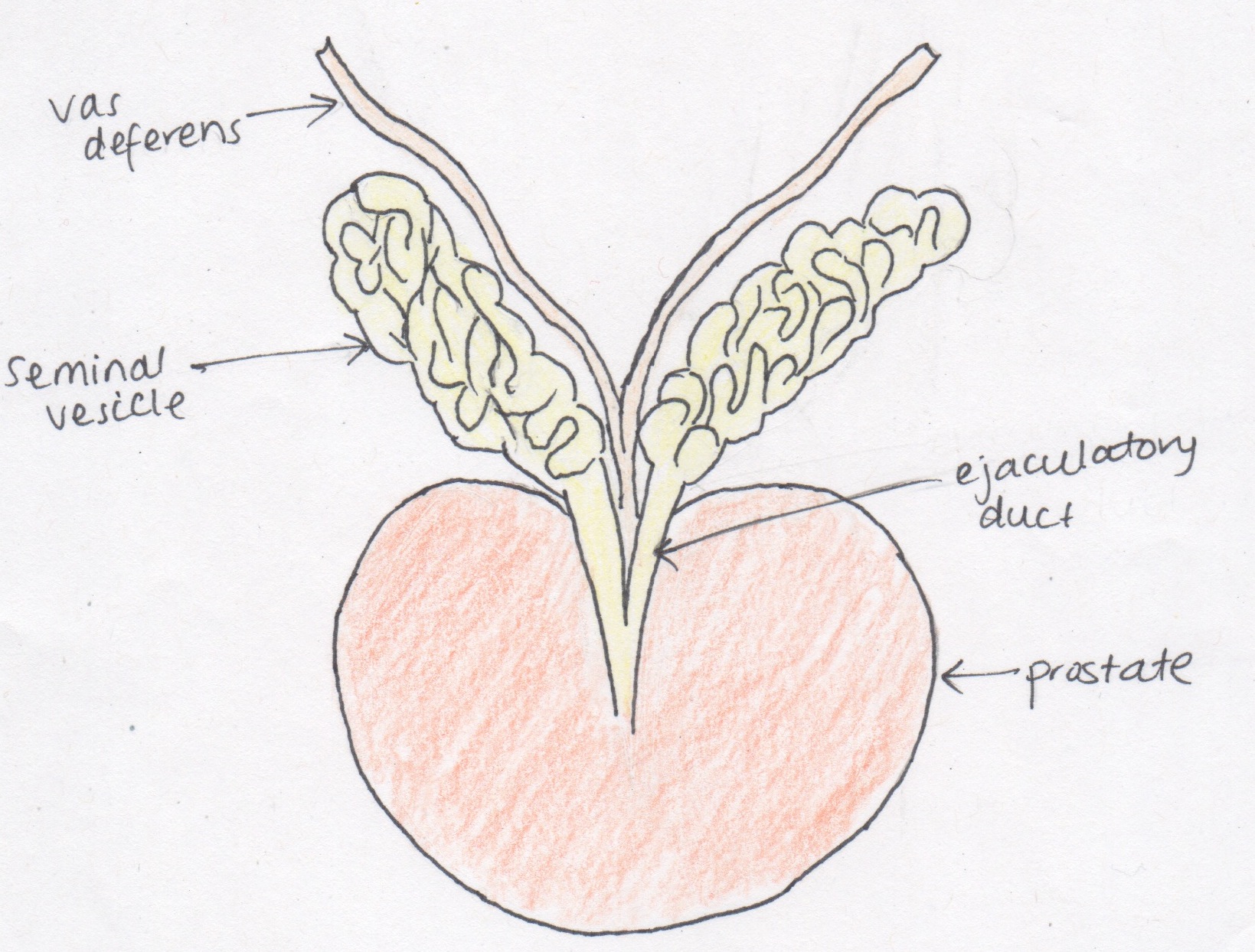
526 KB | This drawing is of the prostate with the seminal vesicles and vas deferens, as first described by Reinier De Graaf, a Dutch anatomists, in 1678. It shows the structures all attaching and places them in their respective positions in relation to the pros... | 1 |
| 13:55, 24 October 2014 | Prepuce.jpeg (file) |  |
382 KB | A summary the steps of prepuce development as described by Hunter in 1935. The drawing resemble and illustrates the observations of the foetal penis at different gestational, depicting that the prepuce is a gradually growing structure during embryogene... | 1 |
| 00:05, 24 October 2014 | Mullerian ducts development.jpeg (file) |  |
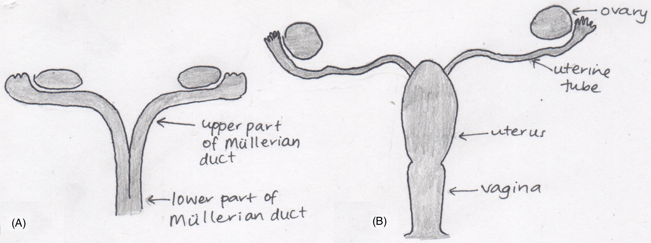
431 KB | This is a diagram illustrating the origin of the female genital organs from the Mullerian ducts, in accordance with the anatomists of the 17th and 18th century. The Mullerian ducts (A) are separated into an upper and lower part, both developing into th... | 2 |
| 16:29, 23 October 2014 | Keibel's model of urogenital organs.jpg (file) |  |
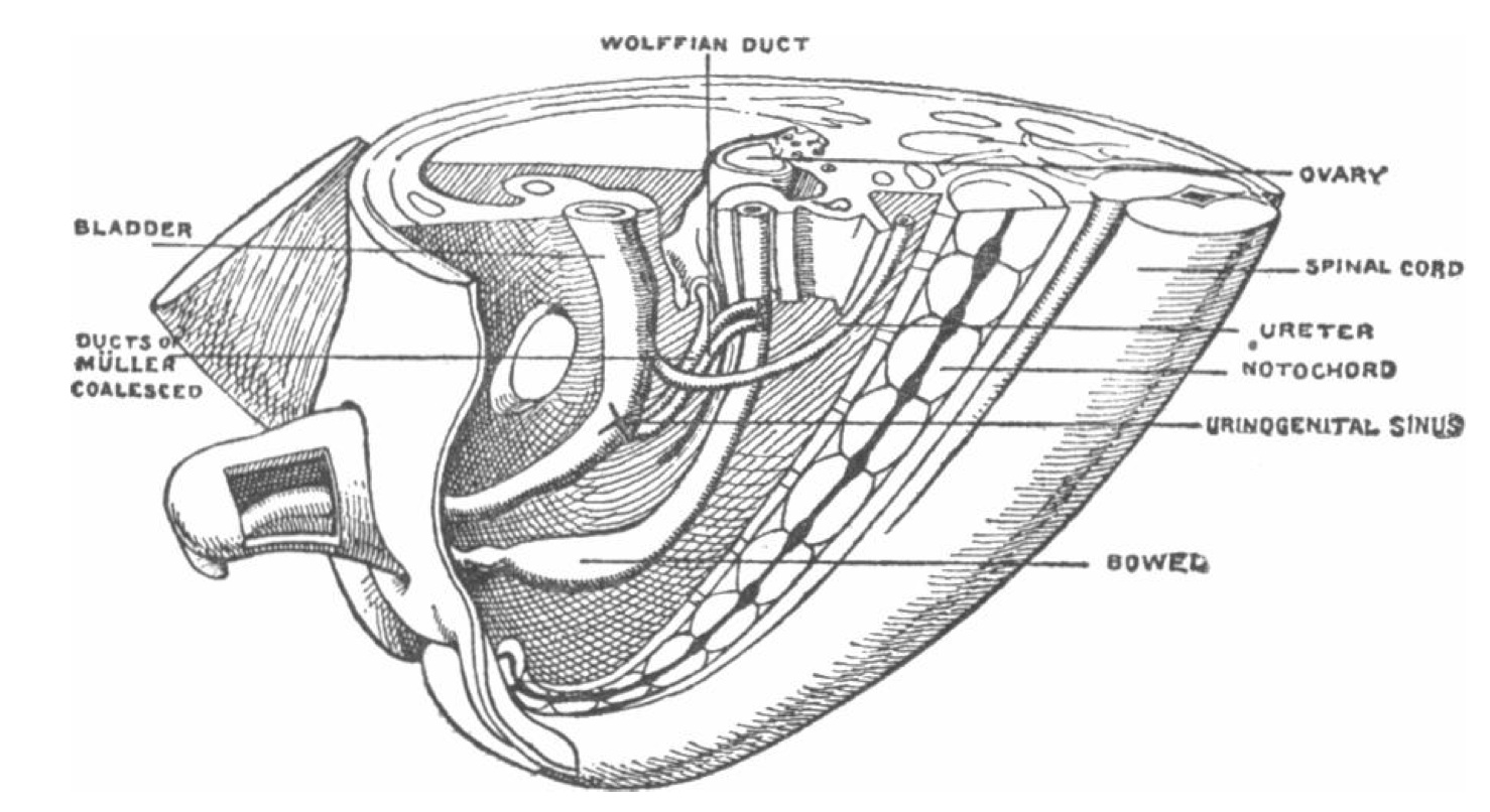
265 KB | This model by the German anatomists and embryologists Franz Keibel marks the location of the later developed hymen, the Mullerian eminence, with an X, at the junction of the urogenital sinus and the Mullerian ducts. It also contains the Mullerian ducts... | 1 |
| 18:51, 19 August 2014 | WNT4 screening in the testis of the tammar wallaby.jpeg (file) |  |
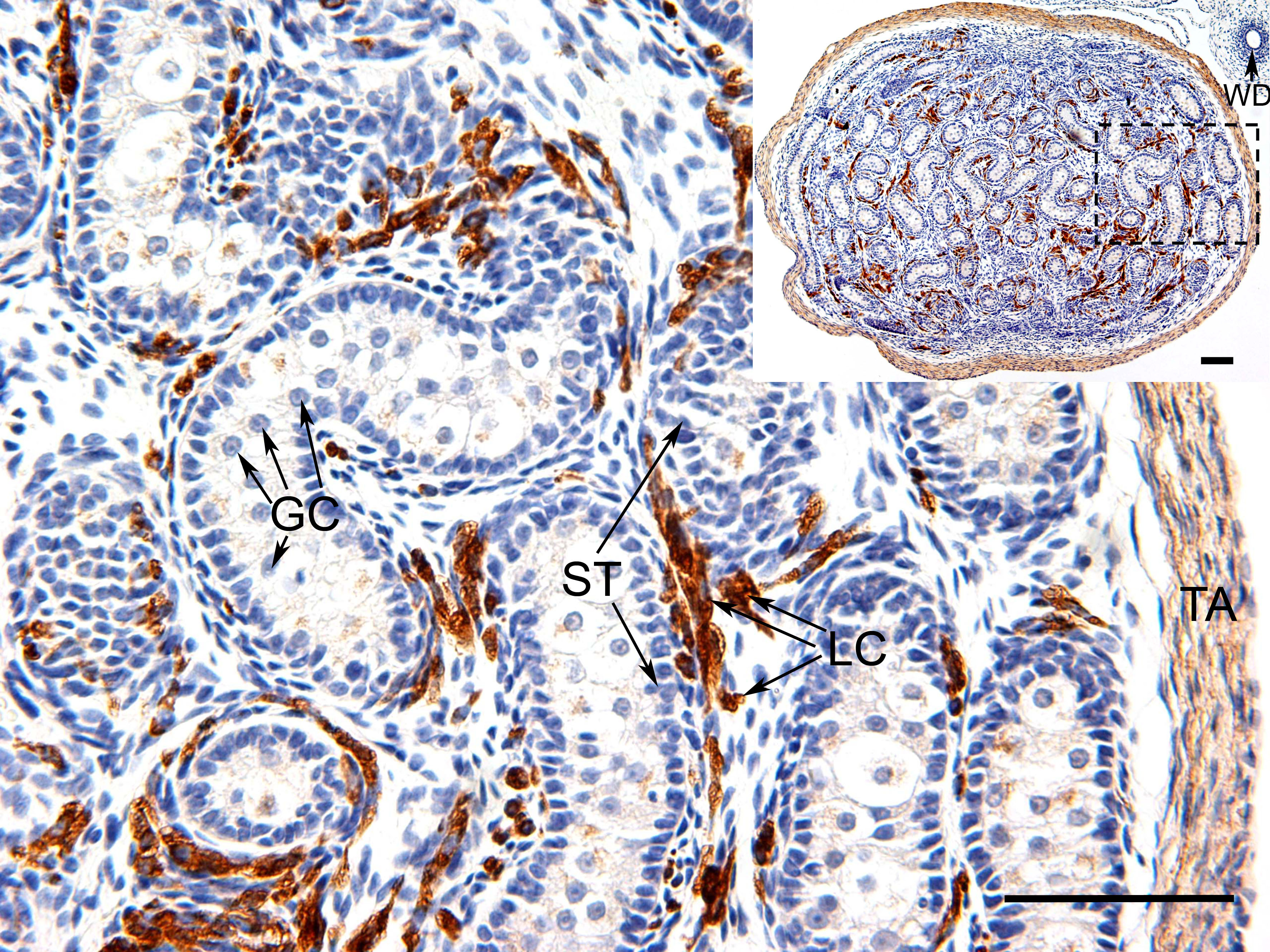
1.91 MB | ==WNT4 screening in the testis of the tammar wallaby== This is an image of a histological slide of the testis of a tammar wallaby treated with WNT4 immunostaining. The brown colouration represents the localisation of the WNT4 protein found within the L... | 1 |