File:ZScan Lines.jpg
Original file (985 × 974 pixels, file size: 322 KB, MIME type: image/jpeg)
Explanation of Diagram
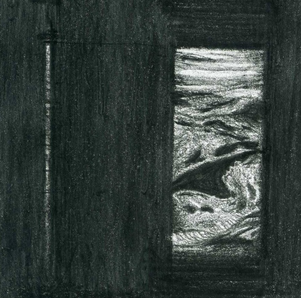
This diagram is designed to show that a single pulse of ultrasound produces one scan line of information, so many pulses of ultrasound together produce many scan lines and ultimately a more complete cross-sectional image of the tissue being examined. A single scan line is shown on the left, and a more complete ultrasound image on the right.
The single scan line is the result of a single ultrasound pulse being emitted from a transducer into tissues, and the echoes of that pulse being received back and interpreted by the ultrasound machine software. The scan line shows light and dark dots that correspond to tissues within the body that have differing material properties.
The image on the right is the combination of many ultrasound pulses and thus many scan lines of information. It shows the profile of a fetus’s head and upper thorax at approximately 17 weeks.
- Note - This image was originally uploaded as part of an undergraduate science student project and may contain inaccuracies in either description or acknowledgements. Students have been advised in writing concerning the reuse of content and may accidentally have misunderstood the original terms of use. If image reuse on this non-commercial educational site infringes your existing copyright, please contact the site editor for immediate removal.
Image Copyright Information
Illustration by z3252833.
Beginning six months after publication, I, z3252833, grant the public the non-exclusive right to copy, distribute, or display the Work under a Creative Commons Attribution-Noncommercial-Share Alike 3.0 Unported license, as described at http://creativecommons.org/licenses/by-nc-sa/3.0/ and http://creativecommons.org/licenses/by-nc-sa/3.0/legalcode.
References
Kremkali, F.W. (2006) Diagnostic Ultrasound Principles and Instruments (7th ed.) St Louis: Saunders Elsevier. pp 3-7
File history
Click on a date/time to view the file as it appeared at that time.
| Date/Time | Thumbnail | Dimensions | User | Comment | |
|---|---|---|---|---|---|
| current | 17:24, 12 September 2010 | | 985 × 974 (322 KB) | Z3252833 (talk | contribs) | ===Explanation of Diagram=== This diagram is designed to show that a single pulse of ultrasound produces one scan line of information, so many pulses of ultrasound together produce many scan lines and ultimately a more complete cross-sectional image of t |
You cannot overwrite this file.
File usage
The following page uses this file: