File:ZDoppler Image of Fetal Aorta.jpg: Difference between revisions
(===What am I looking at?=== This is a colour Doppler ultrasound image depicting a sagittal section of the chest of a fetus. The aortic arch and descending thoracic aorta can be seen extending from the left ventricular outflow tract. Colour is used to ) |
|||
| Line 13: | Line 13: | ||
Author (content provider): Dr. Joe Antony | Author (content provider): Dr. Joe Antony | ||
This image has been reproduced here with the permission of Dr. Joe Antony, who controls the website [http://www.ultrasound-images.com/ Ultrasound Images], "a free gallery of high-resolution, ultrasound, color Doppler and 3D images". Correspondence attesting to this fact will be cheerfully provided upon request. | This image is not classed as a public domain image, but has been reproduced here with the kind permission of Dr. Joe Antony, who controls the website [http://www.ultrasound-images.com/ Ultrasound Images], "a free gallery of high-resolution, ultrasound, color Doppler and 3D images". Correspondence attesting to this fact will be cheerfully provided upon request. | ||
===References=== | ===References=== | ||
<References/> | <References/> | ||
Revision as of 19:00, 10 September 2010
What am I looking at?
This is a colour Doppler ultrasound image depicting a sagittal section of the chest of a fetus. The aortic arch and descending thoracic aorta can be seen extending from the left ventricular outflow tract.
Colour is used to highlight moving structures, providing information about blood flow and associated structures. Colours are generally assigned according to the direction of movement towards or away from the ultrasound beam, with the blue colour indicating movement away from the ultrasound beam and red indicating movement toward the beam.
Image Copyright Information
Image obtained at: http://www.ultrasound-images.com/fetal-chest.htm
Author (content provider): Dr. Joe Antony
This image is not classed as a public domain image, but has been reproduced here with the kind permission of Dr. Joe Antony, who controls the website Ultrasound Images, "a free gallery of high-resolution, ultrasound, color Doppler and 3D images". Correspondence attesting to this fact will be cheerfully provided upon request.
References
File history
Click on a date/time to view the file as it appeared at that time.
| Date/Time | Thumbnail | Dimensions | User | Comment | |
|---|---|---|---|---|---|
| current | 18:56, 10 September 2010 | 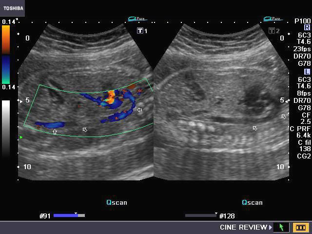 | 640 × 480 (53 KB) | Z3252833 (talk | contribs) | ===What am I looking at?=== This is a colour Doppler ultrasound image depicting a sagittal section of the chest of a fetus. The aortic arch and descending thoracic aorta can be seen extending from the left ventricular outflow tract. Colour is used to |
You cannot overwrite this file.
File usage
The following page uses this file: