File:Z3333865.pax2 and 8.jpg
Original file (1,200 × 1,501 pixels, file size: 339 KB, MIME type: image/jpeg)
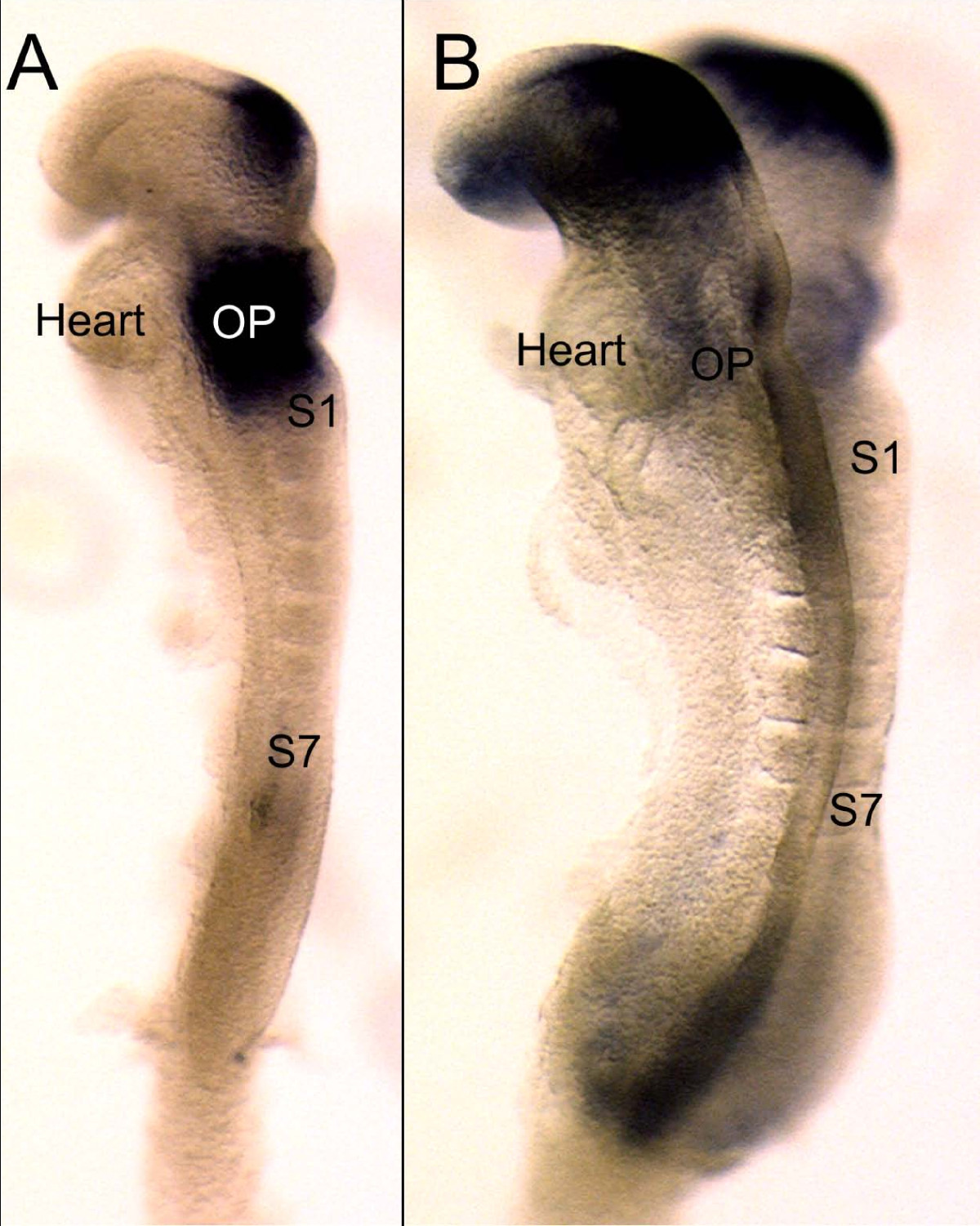
Early expression of Pax2 and Pax8 compared
This image shows the earliest expression of Pax8 (A) and Pax2 (B) in 7 somite (S1, S7) mouse embryos of approximately 8 embryonic days. Note that at this stage Pax8 is more profoundly expressed throughout the area of the future otic placode (OP) just anterior to the first somite (S1). In contrast, Pax2 is more prominently expressed in the brain with only very limited expression in the area of the otic placode (OP). At this stage the heart is anterior to the future otic placode. Bar indicates 100 um.
Reference:<pubmed>20727173</pubmed>
© 2010 Bouchard et al; licensee BioMed Central Ltd. This is an Open Access article distributed under the terms of the Creative Commons Attribution License (http://creativecommons.org/licenses/by/2.0), which permits unrestricted use, distribution, and reproduction in any medium, provided the original work is properly cited.
- Note - This image was originally uploaded as part of an undergraduate science student project and may contain inaccuracies in either description or acknowledgements. Students have been advised in writing concerning the reuse of content and may accidentally have misunderstood the original terms of use. If image reuse on this non-commercial educational site infringes your existing copyright, please contact the site editor for immediate removal.
File history
Click on a date/time to view the file as it appeared at that time.
| Date/Time | Thumbnail | Dimensions | User | Comment | |
|---|---|---|---|---|---|
| current | 13:29, 11 September 2012 | | 1,200 × 1,501 (339 KB) | Z3333865 (talk | contribs) | '''Early expression of Pax2 and Pax8 compared''' This image shows the earliest expression of Pax8 (A) and Pax2 (B) in 7 somite (S1, S7) mouse embryos of approximately 8 embryonic days. Note that at this stage Pax8 is more profoundly expressed throughout |
You cannot overwrite this file.
File usage
The following 2 pages use this file: