File:Week7.jpg: Difference between revisions
From Embryology
No edit summary |
No edit summary |
||
| Line 2: | Line 2: | ||
Image is self drawn by Student based on the diagram and descriptions provided by: <pubmed>15454774</pubmed> | Image is self drawn by Student based on the diagram and descriptions provided by: <pubmed>15454774</pubmed> | ||
{{Template:Student Image}} | |||
Latest revision as of 02:56, 3 October 2012
A diagram of the sagittal section of a week 7 embryo showing the final stages of the development of the sensory structures.
Image is self drawn by Student based on the diagram and descriptions provided by: <pubmed>15454774</pubmed>
- Note - This image was originally uploaded as part of an undergraduate science student project and may contain inaccuracies in either description or acknowledgements. Students have been advised in writing concerning the reuse of content and may accidentally have misunderstood the original terms of use. If image reuse on this non-commercial educational site infringes your existing copyright, please contact the site editor for immediate removal.
File history
Click on a date/time to view the file as it appeared at that time.
| Date/Time | Thumbnail | Dimensions | User | Comment | |
|---|---|---|---|---|---|
| current | 02:50, 3 October 2012 | 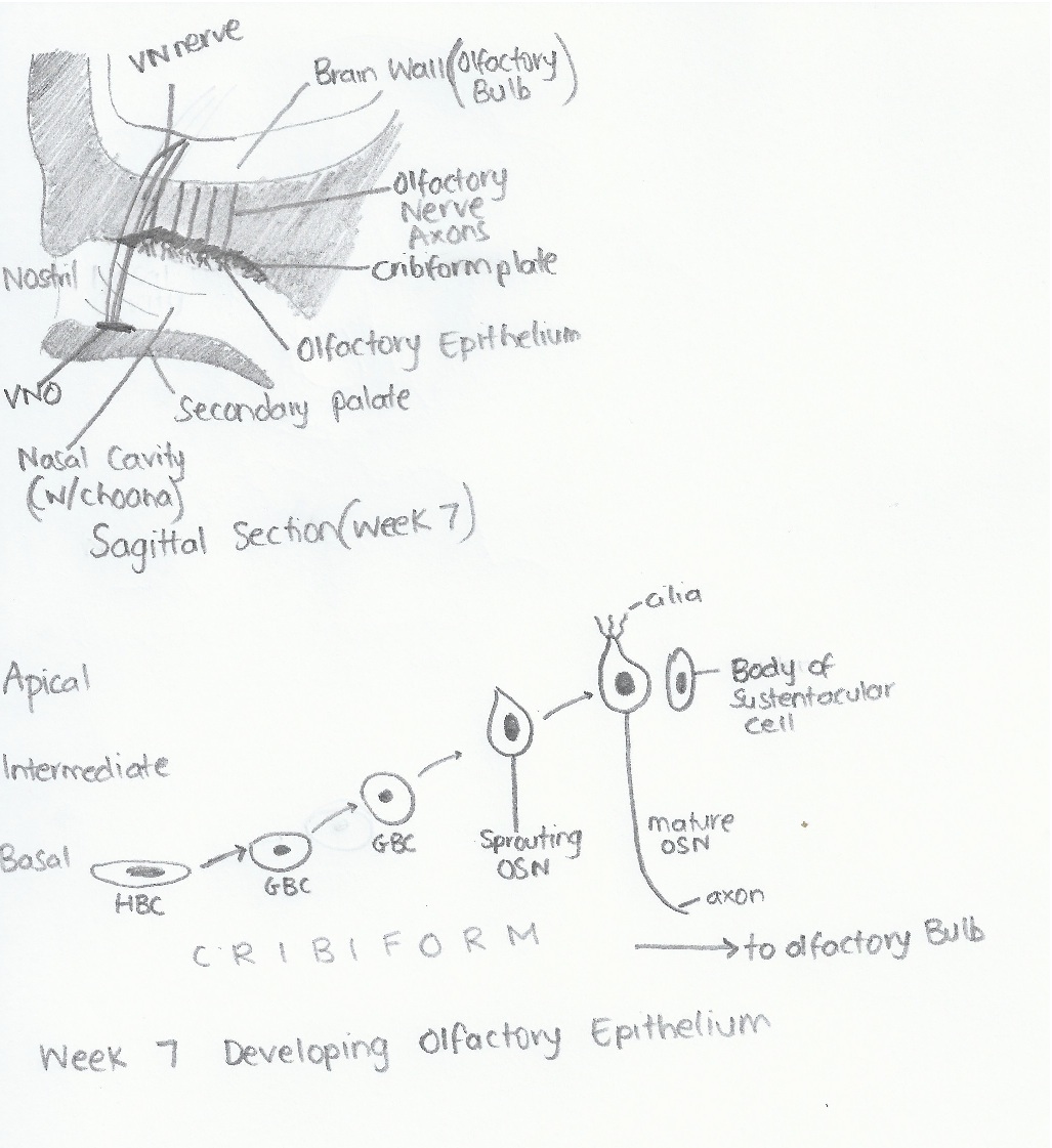 | 1,027 × 1,121 (217 KB) | Z3331264 (talk | contribs) |
You cannot overwrite this file.
File usage
The following 2 pages use this file: