File:WNT4 screening in the testis of the tammar wallaby.jpeg
Original file (3,543 × 2,657 pixels, file size: 1.91 MB, MIME type: image/jpeg)
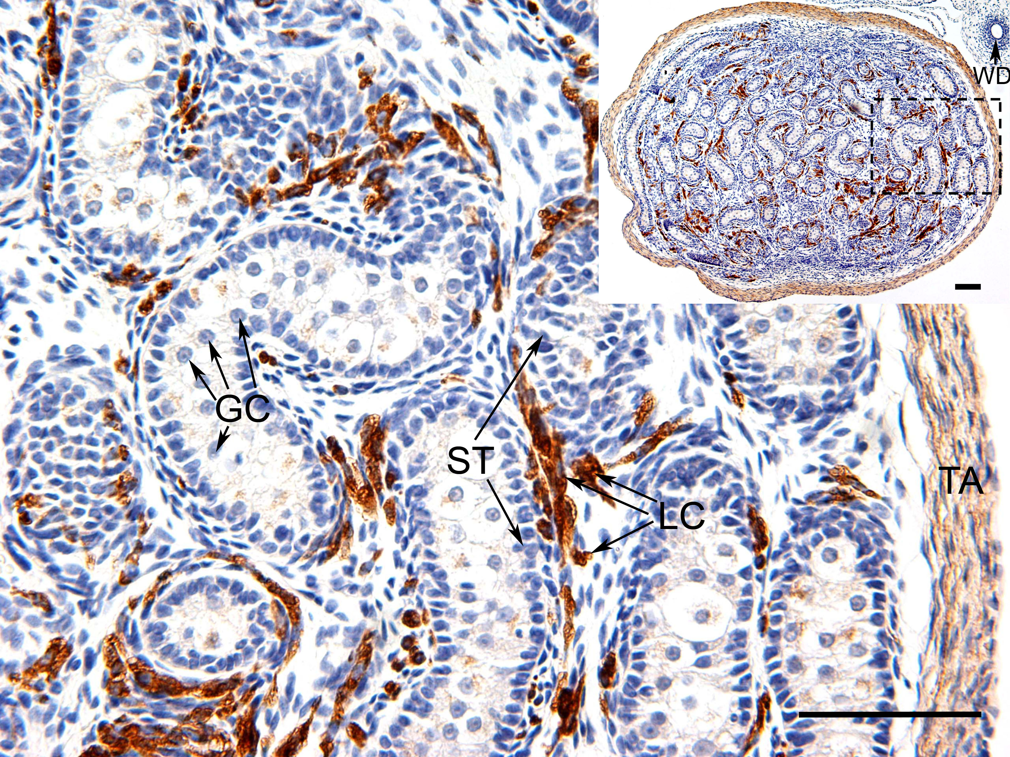
WNT4 screening in the testis of the tammar wallaby
This is an image of a histological slide of the testis of a tammar wallaby treated with WNT4 immunostaining. The brown colouration represents the localisation of the WNT4 protein found within the Leydig cells (LC) and the tunica albuginea (TA). The image also indicates that the germ cells (GC), Sertoli cells (ST) and the Wolffian duct (WD) lack WNT4.
Reference
<pubmed>17014734</pubmed>| Biomed Central
© 2006 Yu et al; licensee BioMed Central Ltd. This is an Open Access article distributed under the terms of the Creative Commons Attribution License, which permits unrestricted use, distribution, and reproduction in any medium, provided the original work is properly cited.
--Mark Hill This is fine and description is good. I have fixed the reference formatting here, that forma is how it should appear on your personal page.
- Note - This image was originally uploaded as part of an undergraduate science student project and may contain inaccuracies in either description or acknowledgements. Students have been advised in writing concerning the reuse of content and may accidentally have misunderstood the original terms of use. If image reuse on this non-commercial educational site infringes your existing copyright, please contact the site editor for immediate removal.
File history
Click on a date/time to view the file as it appeared at that time.
| Date/Time | Thumbnail | Dimensions | User | Comment | |
|---|---|---|---|---|---|
| current | 18:51, 19 August 2014 | | 3,543 × 2,657 (1.91 MB) | Z3415716 (talk | contribs) | ==WNT4 screening in the testis of the tammar wallaby== This is an image of a histological slide of the testis of a tammar wallaby treated with WNT4 immunostaining. The brown colouration represents the localisation of the WNT4 protein found within the L... |
You cannot overwrite this file.
File usage
The following page uses this file: