File:Ventricular Septal Defect (VSD).jpeg: Difference between revisions
No edit summary |
No edit summary |
||
| Line 1: | Line 1: | ||
Description | ===Description=== | ||
''Figure 19 | |||
'' | |||
The view from the left ventricle in this specimen shows a ventricular septal defect with exclusively muscular borders opening towards the outlet of the right ventricle, but with postero-caudal deviation of the muscular outlet septum (yellow dots), causing subaortic stenosis. | The view from the left ventricle in this specimen shows a ventricular septal defect with exclusively muscular borders opening towards the outlet of the right ventricle, but with postero-caudal deviation of the muscular outlet septum (yellow dots), causing subaortic stenosis. | ||
Reference | ===Reference=== | ||
Diane E Spicer, Hao H Hsu, Jennifer Co-Vu, Robert H Anderson, F Jay Fricker | Diane E Spicer, Hao H Hsu, Jennifer Co-Vu, Robert H Anderson, F Jay Fricker | ||
Ventricular septal defect. | Ventricular septal defect. | ||
Orphanet J Rare Dis: 2014, 9;144 | Orphanet J Rare Dis: 2014, 9;144 | ||
Copyright | ===Copyright=== | ||
The open access articles published in BioMed Central's journals are made available under the Creative Commons Attribution (CC-BY) license, which means they are accessible online without any restrictions and can be re-used in any way, subject only to proper attribution (which, in an academic context, usually means citation). | |||
{{Template:Student Image}} | |||
.jpeg&oldid=312964)
.jpeg&action=edit&oldid=312964)
.jpeg&diff=prev&oldid=312964)
.jpeg&oldid=315826)
.jpeg&action=edit&oldid=315826)
.jpeg&diff=next&oldid=315826)
Revision as of 22:19, 25 October 2017
Description
Figure 19
The view from the left ventricle in this specimen shows a ventricular septal defect with exclusively muscular borders opening towards the outlet of the right ventricle, but with postero-caudal deviation of the muscular outlet septum (yellow dots), causing subaortic stenosis.
Reference
Diane E Spicer, Hao H Hsu, Jennifer Co-Vu, Robert H Anderson, F Jay Fricker Ventricular septal defect. Orphanet J Rare Dis: 2014, 9;144
Copyright
The open access articles published in BioMed Central's journals are made available under the Creative Commons Attribution (CC-BY) license, which means they are accessible online without any restrictions and can be re-used in any way, subject only to proper attribution (which, in an academic context, usually means citation).
- Note - This image was originally uploaded as part of an undergraduate science student project and may contain inaccuracies in either description or acknowledgements. Students have been advised in writing concerning the reuse of content and may accidentally have misunderstood the original terms of use. If image reuse on this non-commercial educational site infringes your existing copyright, please contact the site editor for immediate removal.
File history
Click on a date/time to view the file as it appeared at that time.
| Date/Time | Thumbnail | Dimensions | User | Comment | |
|---|---|---|---|---|---|
| current | 13:43, 21 October 2017 | 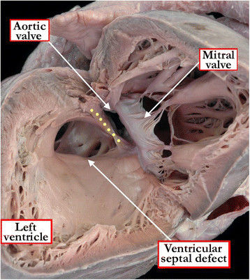 | 358 × 402 (46 KB) | Z5059996 (talk | contribs) | The view from the left ventricle in this specimen shows a ventricular septal defect with exclusively muscular borders opening towards the outlet of the right ventricle, but with postero-caudal deviation of the muscular outlet septum (yellow dots), caus... |
You cannot overwrite this file.
File usage
The following 2 pages use this file:
.jpeg&oldid=315826)