File:Typical tbx protein structure.png
Typical_tbx_protein_structure.png (773 × 164 pixels, file size: 5 KB, MIME type: image/png)
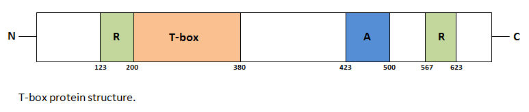
Example using Tbx3 which includes: a T-box DNA-binding domain (orange), R = repression domains (green), A = activation domain (blue) and other protein coding regions (white). The amino and carboxy termini of the protein are labelled N and C respectively. The T-box domain spans 180-200 amino acid residues and is conserved in all T-box proteins. A T-box protein is typically 50 - 78 kDa, within which the T-box domain is typically 17 - 26 kDa. This T-box is conserved in all T-box proteins and spans 180-200 amino acid residues and binds DNA in a sequence-specific manner. They bind to the DNA consensus sequence TCACACCT.
Student drawn image
File history
Click on a date/time to view the file as it appeared at that time.
| Date/Time | Thumbnail | Dimensions | User | Comment | |
|---|---|---|---|---|---|
| current | 16:26, 19 October 2016 | 773 × 164 (5 KB) | Z5020373 (talk | contribs) | Example using Tbx3 which includes: a T-box DNA-binding domain (orange), R = repression domains (green), A = activation domain (blue) and other protein coding regions (white). The amino and carboxy termini of the protein are labelled N and C respective... |
You cannot overwrite this file.
File usage
The following 2 pages use this file: