File:ThyroidDevelopment.png
From Embryology
Size of this preview: 800 × 486 pixels. Other resolution: 1,996 × 1,212 pixels.
Original file (1,996 × 1,212 pixels, file size: 49 KB, MIME type: image/png)
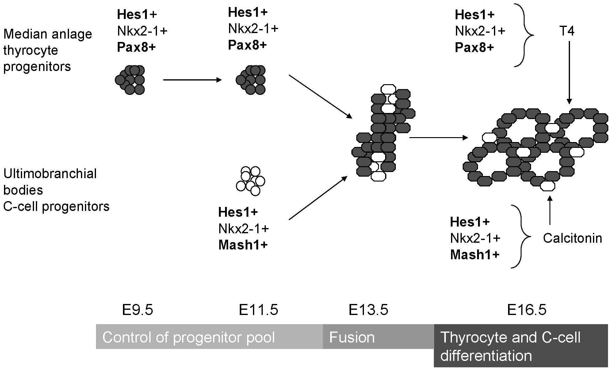
Graphical representation of the endodermal and mesodermal contribution of the thyroid gland
This image summarises the endodermal and mesodermal contribution to the development of the thyroid gland. The progenitor cells are from anterior endoderm and receives buds from left and right endodermal buds covered in mesoderm.
References
<pubmed>21364918</pubmed>
Copyright
Copyright: © 2011 Carre et al. This is an open-access article distributed under the terms of the Creative Commons Attribution License, which permits unrestricted use, distribution, and reproduction in any medium, provided the original author and source are credited.
- Note - This image was originally uploaded as part of an undergraduate science student project and may contain inaccuracies in either description or acknowledgements. Students have been advised in writing concerning the reuse of content and may accidentally have misunderstood the original terms of use. If image reuse on this non-commercial educational site infringes your existing copyright, please contact the site editor for immediate removal.
File history
Click on a date/time to view the file as it appeared at that time.
| Date/Time | Thumbnail | Dimensions | User | Comment | |
|---|---|---|---|---|---|
| current | 13:17, 17 September 2014 | | 1,996 × 1,212 (49 KB) | Z3414648 (talk | contribs) | This image summarises the endodermal and mesodermal contribution to the development of the thyroid gland. The progenitor cells are from anterior endoderm and receives buds from left and right endodermal buds covered in mesoderm. <ref name="PMID10.1371/... |
You cannot overwrite this file.
File usage
The following 2 pages use this file: