File:Stage10 SEM1.jpg
Stage10_SEM1.jpg (277 × 450 pixels, file size: 28 KB, MIME type: image/jpeg)
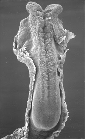
Carnegie Stages 10, 4-5 somites
MH - This description requires checking
Facts: Week 4, 22 - 23 days, 2 - 3.5 mm, Somite Number 4 - 12
View: This is a dorsal view of the embryo. Amniotic membrane removed.
Features: Somite Number 4 - 12, rostral neuropore, neural folds in region of developing brain, neural tube, somites, caudal neuropore, neural fold fuses, remnant of amniotic sac
Identify: rostral neuropore, neural folds in region of developing brain, neural tube, somites (note the different number formed), caudal neuropore, neural fold fuses, cut edge of amniotic sac
Events Ectoderm: Neural fold deeepens, edges approach midline, neural fold fuses, neural plate folds ventrally in brain region
Mesoderm: Somitogenesis, continued segmentation of paraxial mesoderm (4 - 12 somite pairs)
Image Source: Scanning electron micrographs of the Carnegie stages of the early human embryos are reproduced with the permission of Prof Kathy Sulik, from embryos collected by Dr. Vekemans and Tania Attié-Bitach. Images are for educational purposes only and cannot be reproduced electronically or in writing without permission.
File history
Click on a date/time to view the file as it appeared at that time.
| Date/Time | Thumbnail | Dimensions | User | Comment | |
|---|---|---|---|---|---|
| current | 15:32, 10 August 2009 | | 277 × 450 (28 KB) | MarkHill (talk | contribs) | Carnegie Stages 10, 4-5 somites Features: brain fold, neural groove, amniotic sac, presomitic mesoderm, embryonic disc, primitive node, primative streak, primative groove, connecting stalk Facts: Week 3, 21 days, 4 - 5 somites, View: Dorsal view amnioti |
You cannot overwrite this file.
File usage
The following 11 pages use this file: