File:SpinabifidaMeningocele1.jpg
From Embryology
No higher resolution available.
SpinabifidaMeningocele1.jpg (674 × 277 pixels, file size: 32 KB, MIME type: image/jpeg)
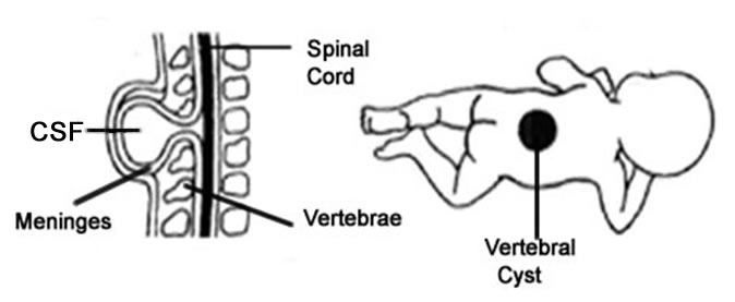
SpinabifidaMeningocele.jpg (674 × 277 pixels, file size: 32 KB, MIME type: image/jpeg) Modified image showing Spina Bifida Meningocele characterized by normal spinal cord, divided outer vertebrae and meninges surrounding the spinal cord protruding from the divided vertebrae as a cyst.
File history
Click on a date/time to view the file as it appeared at that time.
| Date/Time | Thumbnail | Dimensions | User | Comment | |
|---|---|---|---|---|---|
| current | 13:33, 18 September 2009 | 674 × 277 (32 KB) | Z3187802 (talk | contribs) | SpinabifidaMeningocele.jpg (674 × 277 pixels, file size: 32 KB, MIME type: image/jpeg) Modified image showing Spina Bifida Meningocele characterized by normal spinal cord, divided outer vertebrae and meninges surrounding the spinal cord protruding fro |
You cannot overwrite this file.
File usage
The following page uses this file: