File:Skeletal muscle structure.jpg
Skeletal_muscle_structure.jpg (754 × 535 pixels, file size: 99 KB, MIME type: image/jpeg)
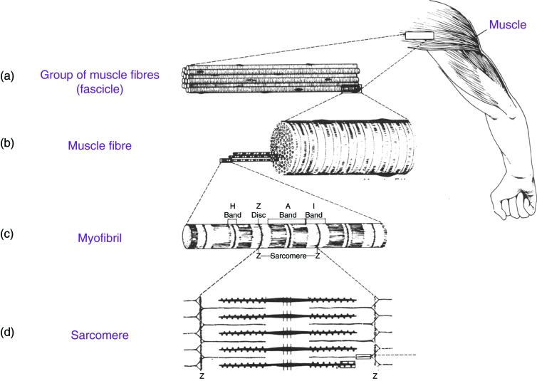
Diagram of a striated muscle (a) Group of muscle fibers make up a fascicle (b) A muscle fiber is made of thousands of myofibrils (c) Myofibrils contain dark (A) and light (I) bands and Z bands where actin originates from and moves towards the A band. (d) Myofibrils are made of repeating units known as sarcomeres. Sarcomeres contains thin actin filaments and thick myosin filaments.
References
- ↑ <pubmed>PMC3963759</pubmed>
Copyright © 2013 AL-Khayat, licensee Bloomsbury Qatar Foundation Journals. This is an open access article distributed under the terms of the Creative Commons Attribution license CC BY 3.0, which permits unrestricted use, distribution and reproduction in any medium, provided the original work is properly cited.
File history
Click on a date/time to view the file as it appeared at that time.
| Date/Time | Thumbnail | Dimensions | User | Comment | |
|---|---|---|---|---|---|
| current | 23:22, 22 October 2014 | | 754 × 535 (99 KB) | Z3418989 (talk | contribs) | Diagram of a striated muscle (a) Group of muscle fibers make up a fascicle (b) A muscle fiber is made of thousands of myofibrils (c) Myofibrils contain dark (A) and light (I) bands and Z bands where actin originates from and moves towards the A band.... |
You cannot overwrite this file.
File usage
The following 2 pages use this file: