File:Semilobar holoprosencephaly.jpg

Original file (1,024 × 1,024 pixels, file size: 54 KB, MIME type: image/jpeg)
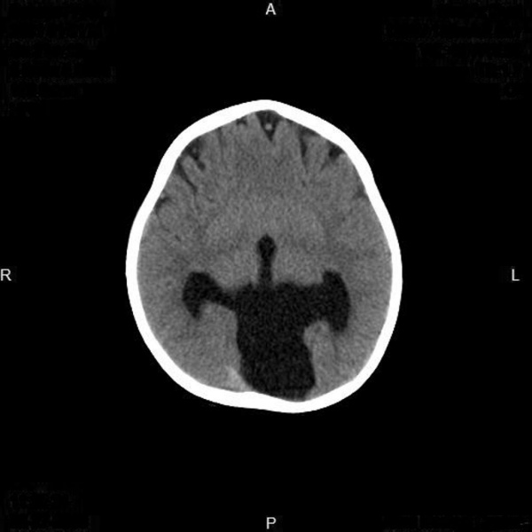
MRI depicting a transverse section of the brain, with the hemispheres of the fore brain partially fused, known as semilobar holoprosencephaly.
Copyright
Creative Commons License
You don't need to ask permission to use these images provided:
- you attribute the contributing user
- the use is non-commercial
- you do not copyright the material
Attribution
Case courtesy of Dr Ruslan Esedov, https://radiopaedia.org/, Radiopaedia.org. From the case https://radiopaedia.org/cases/7504 rID: 7504
Reference
Agrawal, R. (2016). Holoprosencephaly | Radiology Reference Article | Radiopaedia.org. [online] Radiopaedia.org. Available at: https://radiopaedia.org/articles/holoprosencephaly.
- Note - This image was originally uploaded as part of an undergraduate science student project and may contain inaccuracies in either description or acknowledgements. Students have been advised in writing concerning the reuse of content and may accidentally have misunderstood the original terms of use. If image reuse on this non-commercial educational site infringes your existing copyright, please contact the site editor for immediate removal.
File history
Click on a date/time to view the file as it appeared at that time.
| Date/Time | Thumbnail | Dimensions | User | Comment | |
|---|---|---|---|---|---|
| current | 12:59, 22 October 2016 |  | 1,024 × 1,024 (54 KB) | Z5019880 (talk | contribs) |
You cannot overwrite this file.
File usage
The following 2 pages use this file: