File:Scans of Supravalvular Aortic Stenosis and Pulmonary Stenosis.jpg
Scans_of_Supravalvular_Aortic_Stenosis_and_Pulmonary_Stenosis.jpg (665 × 436 pixels, file size: 114 KB, MIME type: image/jpeg)
original file name: Kjr-11-4-g009.jpg
http://www.ncbi.nlm.nih.gov/pmc/articles/PMC2799649/
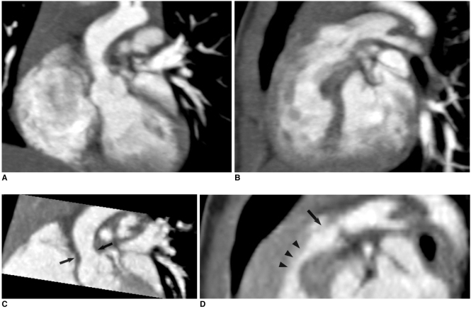
Fig. 9 Combo CT scan comprised of non-ECG-synchronized spiral scan with usual scan range (A, B) and prospective ECG-triggered sequential scan with narrow scan range confined to conotruncal area of heart (C, D) in 9-months-old boy with Williams syndrome. Supravalvular aortic stenosis (arrows on C) and combined valvar (arrow on D) and subvalvar (arrowheads on D) pulmonary stenoses are clearly shown on prospective ECG-triggered sequential CT images (C, D). Dose estimates are 1.6 mSv for non-ECG-synchronized spiral scan and 0.2 mSv for prospective ECG-triggered sequential scan.
Copyright © 2010 The Korean Society of Radiology. This is an Open Access article distributed under the terms of the Creative Commons Attribution Non-Commercial License (http://creativecommons.org/licenses/by-nc/3.0) which permits unrestricted non-commercial use, distribution, and reproduction in any medium, provided the original work is properly cited.
File history
Click on a date/time to view the file as it appeared at that time.
| Date/Time | Thumbnail | Dimensions | User | Comment | |
|---|---|---|---|---|---|
| current | 14:52, 17 September 2011 |  | 665 × 436 (114 KB) | Z3331556 (talk | contribs) | original file name: Kjr-11-4-g009.jpg http://www.ncbi.nlm.nih.gov/pmc/articles/PMC2799649/ Fig. 9 Combo CT scan comprised of non-ECG-synchronized spiral scan with usual scan range (A, B) and prospective ECG-triggered sequential scan with narrow scan ran |
You cannot overwrite this file.
File usage
The following 2 pages use this file:
