File:Retinal Disc to form Optic Cup at Carnegie stage 14.jpg: Difference between revisions
From Embryology
(At Carnegie stage 14, the retinal disc becomes invaginated to form the optic cup. Lens Pit can also be seen forming. Contour forming slowly. Based upon images by: R O'Rahilly The prenatal development of the human eye. Exp. Eye Res.: 1975, 21(2);93-112 F) |
(No difference)
|
Latest revision as of 21:11, 4 October 2012
At Carnegie stage 14, the retinal disc becomes invaginated to form the optic cup. Lens Pit can also be seen forming. Contour forming slowly.
Based upon images by: R O'Rahilly The prenatal development of the human eye. Exp. Eye Res.: 1975, 21(2);93-112 Figure 3.
Beginning six months after publication, I, (z3374173) grant the public the non-exclusive right to copy, distribute, or display the Work under a Creative Commons Attribution-Noncommercial-Share Alike 3.0 Unported license, as described at http://creativecommons.org/licenses/by-nc-sa/3.0/ and http://creativecommons.org/licenses/by-nc-sa/3.0/legalcode.
File history
Click on a date/time to view the file as it appeared at that time.
| Date/Time | Thumbnail | Dimensions | User | Comment | |
|---|---|---|---|---|---|
| current | 21:11, 4 October 2012 | 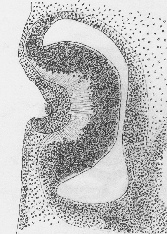 | 572 × 800 (400 KB) | Z3374173 (talk | contribs) | At Carnegie stage 14, the retinal disc becomes invaginated to form the optic cup. Lens Pit can also be seen forming. Contour forming slowly. Based upon images by: R O'Rahilly The prenatal development of the human eye. Exp. Eye Res.: 1975, 21(2);93-112 F |
You cannot overwrite this file.
File usage
The following 2 pages use this file: