File:Pone.0015329.g001 z5039628.jpg
Pone.0015329.g001_z5039628.jpg (483 × 388 pixels, file size: 135 KB, MIME type: image/jpeg)
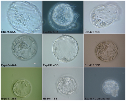
Picture Description: Photographic examples representing the different scores of blastocysts donated for stem cell derivation.
HS475. Fully hatched blastocyst with clear tightly packed ICM. This embryo generated a new hESC line, HS475. Exp462. Hatching blastocyst with average number of cells in ICM, even trophectoderm layer. Exp472. Hatching blastocyst. Trophectoderm layer is uneven and the cells in the ICM are few in number and loosely packed. Exp404. Fully expanded blastocyst with clear tightly packed ICM and even trophectoderm layer. Exp439. Fully expanded blastocyst, with thin zona pellucida. Small ICM and trophectoderm cells of different size. Exp412. Fully developed blastocoel. Zona pellucida is still thick, meaning that the blastocyst is not fully expanded. Exp367. Early blastocyst. HS361. Early blastocyst with blastocoels filling less than 50% of embryo volume. This embryo generated the hESC line HS361. Exp457. This embryo is still compacted and has not reached blastocyst stage. All pictures were taken with a 40x objective, phase contrast or DIC, scale bar 50 µm. (Exp = experiment number. HS is used as a prefix for established hESC lines at Karolinska Institutet).
Picture Link: https://www.ncbi.nlm.nih.gov/pmc/articles/PMC3013107/figure/pone-0015329-g001/
Article Title: No Relationship between Embryo Morphology and Successful Derivation of Human Embryonic Stem Cell Lines
Article Authors: Susanne Ström, Kenny Rodriguez-Wallberg, Frida Holm, Rosita Bergström, Linda Eklund, Anne-Marie Strömberg, and Outi Hovatta
Article Link: https://www.ncbi.nlm.nih.gov/pmc/articles/PMC3013107/
Published online 2010 Dec 31. doi: 10.1371/journal.pone.0015329
PMC copyright statement: Copyright Ström et al. This is an open-access article distributed under the terms of the Creative Commons Attribution License, which permits unrestricted use, distribution, and reproduction in any medium, provided the original author and source are credited.
| Mark Hill 29 August 2016 - Here is the correct reference citation. If you Google the paper title you want the PMC link and from there you can find the PMID. If you continue to have problems, just a see me.
|
- Note - This image was originally uploaded as part of an undergraduate science student project and may contain inaccuracies in either description or acknowledgements. Students have been advised in writing concerning the reuse of content and may accidentally have misunderstood the original terms of use. If image reuse on this non-commercial educational site infringes your existing copyright, please contact the site editor for immediate removal.
File history
Click on a date/time to view the file as it appeared at that time.
| Date/Time | Thumbnail | Dimensions | User | Comment | |
|---|---|---|---|---|---|
| current | 22:21, 21 August 2016 |  | 483 × 388 (135 KB) | Z5039628 (talk | contribs) | Picture Description: Photographic examples representing the different scores of blastocysts donated for stem cell derivation. HS475. Fully hatched blastocyst with clear tightly packed ICM. This embryo generated a new hESC line, HS475. Exp462. Hatching... |
You cannot overwrite this file.
File usage
The following page uses this file:
