File:Picture 1.JPG
From Embryology

Size of this preview: 800 × 285 pixels. Other resolution: 1,593 × 567 pixels.
Original file (1,593 × 567 pixels, file size: 109 KB, MIME type: image/jpeg)
File history
Click on a date/time to view the file as it appeared at that time.
| Date/Time | Thumbnail | Dimensions | User | Comment | |
|---|---|---|---|---|---|
| current | 17:18, 27 August 2009 | 1,593 × 567 (109 KB) | Z3224449 (talk | contribs) | ||
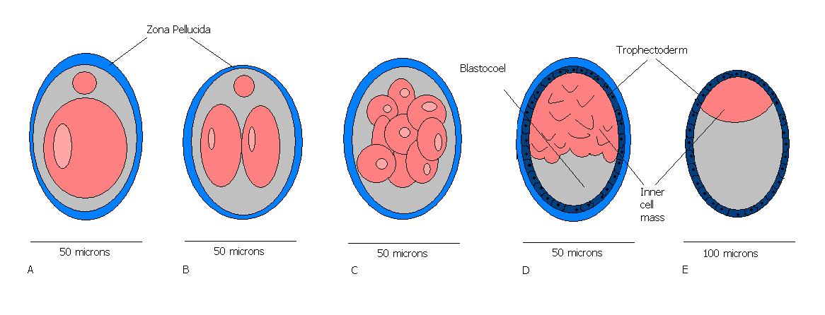
| 12:11, 23 August 2009 | 1,166 × 446 (67 KB) | Z3224449 (talk | contribs) | Figure 1: Illustration of Mouse embryonic stages of development from fertilization to zona free blastocyst (stages 1 to 5). E.Newton 2009 A)Theiler stage 1: one celled embryo B)Theiler stage 2: Dividing embryo ( 2 cells present) C)Theiler stage 3: Morula | ||
| 12:09, 23 August 2009 | 1,166 × 446 (67 KB) | Z3224449 (talk | contribs) |
You cannot overwrite this file.
File usage
The following page uses this file: