File:Pharyngeal arch one and two in mice.png: Difference between revisions
(The image shows the pharyngeal arches one and two in mice embryo. All three layers ectoderm, mesoderm and endoderm from arch one and two contribute to the formation of the ear. <pubmed>22110697</pubmed> Citation: Diman NYS-G, Remacle S, Bertrand N, Pi) |
No edit summary |
||
| Line 1: | Line 1: | ||
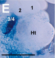
1 and 2 respectively mark the endodermal side of pharyngeal arches one and two and Ht shows where the heart lies in the embryo. | |||
The image shows the pharyngeal arches one and two in mice embryo. All three layers ectoderm, mesoderm and endoderm from arch one and two contribute to the formation of the ear. | The image shows the pharyngeal arches one and two in mice embryo. All three layers ectoderm, mesoderm and endoderm from arch one and two contribute to the formation of the ear. | ||
Revision as of 10:31, 3 October 2012
1 and 2 respectively mark the endodermal side of pharyngeal arches one and two and Ht shows where the heart lies in the embryo.
The image shows the pharyngeal arches one and two in mice embryo. All three layers ectoderm, mesoderm and endoderm from arch one and two contribute to the formation of the ear.
<pubmed>22110697</pubmed>
Citation: Diman NYS-G, Remacle S, Bertrand N, Picard JJ, Zaffran S, et al. (2011) A Retinoic Acid Responsive Hoxa3 Transgene Expressed in Embryonic Pharyngeal Endoderm, Cardiac Neural Crest and a Subdomain of the Second Heart Field. PLoS ONE 6(11): e27624. doi:10.1371/journal.pone.0027624
Copyright: © 2011 Diman et al. This is an open-access article distributed under the terms of the Creative Commons Attribution License, which permits unrestricted use, distribution, and reproduction in any medium, provided the original author and source are credited.
File history
Click on a date/time to view the file as it appeared at that time.
| Date/Time | Thumbnail | Dimensions | User | Comment | |
|---|---|---|---|---|---|
| current | 10:29, 3 October 2012 |  | 448 × 472 (326 KB) | Z3333794 (talk | contribs) | The image shows the pharyngeal arches one and two in mice embryo. All three layers ectoderm, mesoderm and endoderm from arch one and two contribute to the formation of the ear. <pubmed>22110697</pubmed> Citation: Diman NYS-G, Remacle S, Bertrand N, Pi |
You cannot overwrite this file.
File usage
The following 2 pages use this file: