File:Patterns of zona pellucida deposition.JPG

Original file (475 × 711 pixels, file size: 44 KB, MIME type: image/jpeg)
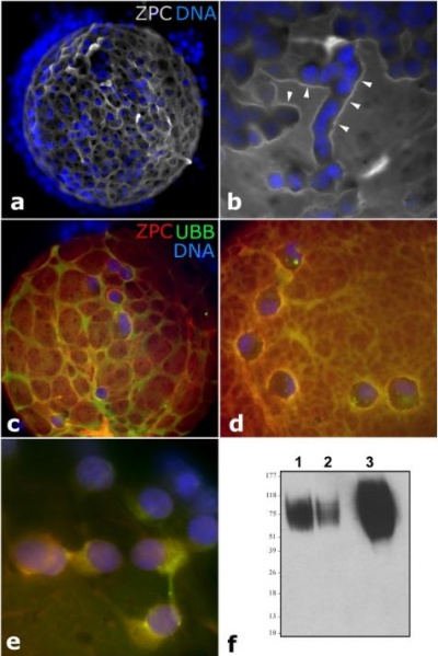
Patterns of Zona Pellucida Deposition
(a, b) Accumulation of ZPC (gray) in ridges (arrowheads) adjacent to zona-adhering corona radiata cells (blue = nuclei stained with DAPI).
(c-e) Colocalization of ZPC (red) and ubiquitin (green) in the zona pellucida (c, d) and in the cytoplasm of corona radiata cells
(e); DNA was counterstained with DAPI (blue). Western blotting of ZPC protein in the isolated cumulus/corona cells from 60 oocyte cumulus complexes (lane 1), in 60 zona-free oocytes (lane 2) and in soluble zona proteins isolated from 60 oocytes (lane 3).
Reference
<pubmed>21383844</pubmed>
Copyright
Zimmerman et al. This is an open-access article distributed under the terms of the Creative Commons Attribution License, which permits unrestricted use, distribution, and reproduction in any medium, provided the original author and source are credited.
File history
Click on a date/time to view the file as it appeared at that time.
| Date/Time | Thumbnail | Dimensions | User | Comment | |
|---|---|---|---|---|---|
| current | 10:54, 25 August 2011 | | 475 × 711 (44 KB) | Z3332824 (talk | contribs) | ==Patterns of zona pellucida deposition== (a, b) Accumulation of ZPC (gray) in ridges (arrowheads) adjacent to zona-adhering corona radiata cells (blue = nuclei stained with DAPI). (c-e) Colocalization of ZPC (red) and ubiquitin (green) in the zona pellu |
You cannot overwrite this file.
File usage
The following page uses this file: