File:Ovarian Structural scematic and stained cortical region of an ovariana stem cell .jpg
From Embryology
Ovarian_Structural_scematic_and_stained_cortical_region_of_an_ovariana_stem_cell_.jpg (600 × 307 pixels, file size: 89 KB, MIME type: image/jpeg)
S13048-015-0184-9-1.jpg
PMID 26250560 [[1]]
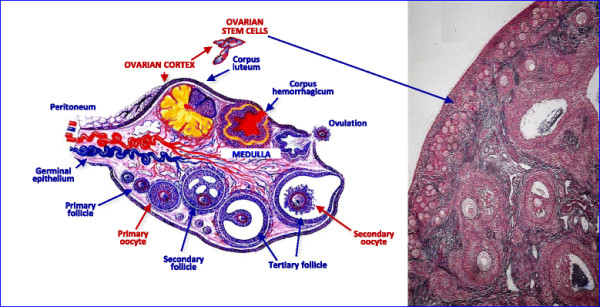
Structure of human ovary. Left – Schematic representation of the ovarian structure in a woman in reproductive age, showing the evolution of primary follicles to corpora lutea that cyclically occur in the cortex of the organ. Right – The ovarian cortex is the presumable site of the ovarian stem cell location in this ovary preparation after hematoxylin/eosin staining
File history
Click on a date/time to view the file as it appeared at that time.
| Date/Time | Thumbnail | Dimensions | User | Comment | |
|---|---|---|---|---|---|
| current | 14:21, 14 August 2015 |  | 600 × 307 (89 KB) | Z5020317 (talk | contribs) | S13048-015-0184-9-1.jpg PMID 26250560 http://www.ovarianresearch.com/content/8/1/55 Structure of human ovary. Left – Schematic representation of the ovarian structure in a woman in reproductive age, showing the evolution of primary follicles to... |
You cannot overwrite this file.
File usage
The following page uses this file:
