File:Normal cochlea.png

Original file (571 × 789 pixels, file size: 141 KB, MIME type: image/png)
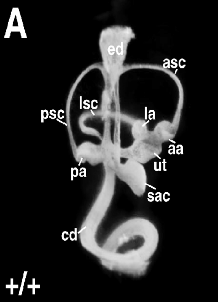
Wild-type inner ear showing normal morphology.
Structures are labeled as follows: aa, anterior ampulla; asc, anterior semicircular canal; cd, cochlea duct; ed, endolymphatic duct; la, lateral ampulla; lsc, lateral semicircular canal; pa, posterior ampulla; psc, posterior semicircular canal; sac, saccule; ut, utricle.
Reference: <pubmed>16410827</pubmed>
Copyright: © 2006 Kiernan et al. This is an open-access article distributed under the terms of the Creative Commons Attribution License, which permits unrestricted use, distribution, and reproduction in any medium, provided the original author and source are credited.
File history
Click on a date/time to view the file as it appeared at that time.
| Date/Time | Thumbnail | Dimensions | User | Comment | |
|---|---|---|---|---|---|
| current | 10:13, 22 August 2012 |  | 571 × 789 (141 KB) | Z3333865 (talk | contribs) | Wild-type inner ear showing normal morphology. Structures are labeled as follows: aa, anterior ampulla; asc, anterior semicircular canal; cd, cochlea duct; ed, endolymphatic duct; la, lateral ampulla; lsc, lateral semicircular canal; pa, posterior ampul |
You cannot overwrite this file.
File usage
The following 2 pages use this file: