File:Neurpore cell shape changes.png
Original file (785 × 1,073 pixels, file size: 1.11 MB, MIME type: image/png)
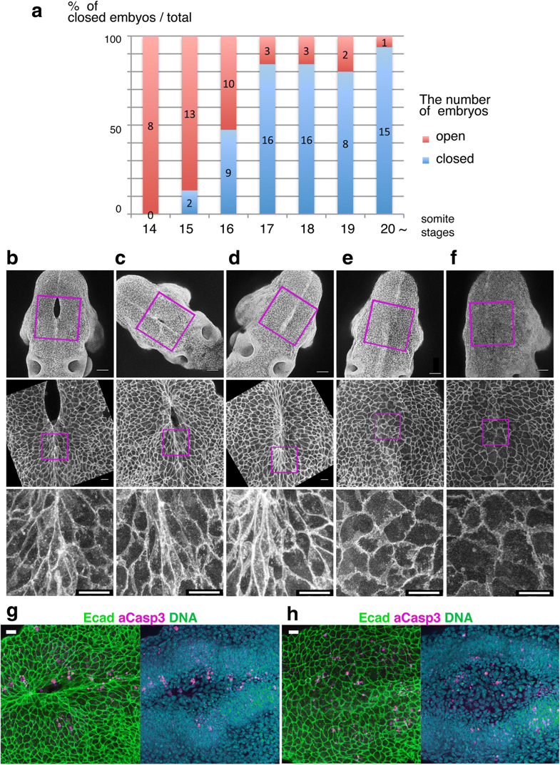
Cell shape changes in the midline after mid-hindbrain neuropore (MHNP) closure in vivo
a The relationship between the closing of MHNP and the somite stage in ICR strain mouse.
b-f Dorsal views of embryos stained with E-cadherin (Ecad). Non-neural ectodermal cells change their shapes dramatically from bipolar to polygonal after the MHNP closure.
g, h Higher manigification views of embryos (c, e) that were co-stained with Ecad, apoptotic marker active-Caspase 3 (aCasp 3), and nuclear staining (DNA). Dorsal views, left is rostral side.
Scale bars: (b-f) 100 μm in upper panels, and 25 μm in (g, h) middle and (b-f) lower panels
Reference
Shinotsuka N, Yamaguchi Y, Nakazato K, Matsumoto Y, Mochizuki A & Miura M. (2018). Caspases and matrix metalloproteases facilitate collective behavior of non-neural ectoderm after hindbrain neuropore closure. BMC Dev. Biol. , 18, 17. PMID: 30064364 DOI.
copyright
© The Author(s). 2018
This article is distributed under the terms of the Creative Commons Attribution 4.0 International License (http://creativecommons.org/licenses/by/4.0/), which permits unrestricted use, distribution, and reproduction in any medium, provided you give appropriate credit to the original author(s) and the source, provide a link to the Creative Commons license, and indicate if changes were made. The Creative Commons Public Domain Dedication waiver (http://creativecommons.org/publicdomain/zero/1.0/) applies to the data made available in this article, unless otherwise stated.
Fig. 3
- Note - This image was originally uploaded as part of an undergraduate science student project and may contain inaccuracies in either description or acknowledgements. Students have been advised in writing concerning the reuse of content and may accidentally have misunderstood the original terms of use. If image reuse on this non-commercial educational site infringes your existing copyright, please contact the site editor for immediate removal.
File history
Click on a date/time to view the file as it appeared at that time.
| Date/Time | Thumbnail | Dimensions | User | Comment | |
|---|---|---|---|---|---|
| current | 11:46, 7 August 2018 | | 785 × 1,073 (1.11 MB) | Z5163485 (talk | contribs) | Fig. 3 Cell shape changes in the midline after mid-hindbrain neuropore (MHNP) closure in vivo. a The relationship between the closing of MHNP and the somite stage in ICR strain mouse. b-f Dorsal views of embryos stained with E-cadherin (Ecad). Non-neur... |
You cannot overwrite this file.
File usage
There are no pages that use this file.