File:Mouse Neural Crest Contributions to Craniofacial Bones.jpg
From Embryology
Mouse_Neural_Crest_Contributions_to_Craniofacial_Bones.jpg (600 × 245 pixels, file size: 30 KB, MIME type: image/jpeg)
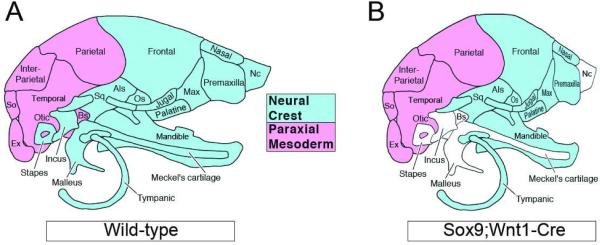
A) Diagram showing the paraxial mesoderm (red) and neural crest (blue) contribution to the mouse head skeleton. Lateral view, anterior to right. B) Sox9 embryo with domed skull, the missing skeletal elements are shown in white.
Reference
Lee YH & Saint-Jeannet JP. (2011). Sox9 function in craniofacial development and disease. Genesis , 49, 200-8. PMID: 21309066 DOI.
Copyright
Copyright © 2011 Wiley‐Liss, Inc.
- Note - This image was originally uploaded as part of an undergraduate science student project and may contain inaccuracies in either description or acknowledgements. Students have been advised in writing concerning the reuse of content and may accidentally have misunderstood the original terms of use. If image reuse on this non-commercial educational site infringes your existing copyright, please contact the site editor for immediate removal.
File history
Click on a date/time to view the file as it appeared at that time.
| Date/Time | Thumbnail | Dimensions | User | Comment | |
|---|---|---|---|---|---|
| current | 14:26, 28 August 2018 | 600 × 245 (30 KB) | Z5160977 (talk | contribs) | A) Diagram showing the paraxial mesoderm (red) and neural crest (blue) contribution to the mouse head skeleton. Lateral view, anterior to right. B) Sox9 embryo with domed skull, the missing skeletal elements are shown in white. |
You cannot overwrite this file.
File usage
There are no pages that use this file.
