File:Heart Tube Fusion Model of Early Development.png

Original file (772 × 1,032 pixels, file size: 301 KB, MIME type: image/png)
<pubmed> PMC2691808 </pubmed>
Received: 9 December 2008 / Accepted: 22 December 2008 / Published online: 30 January 2009 Ó The Author(s) 2009. This article is published with open access at Springerlink.com
Description
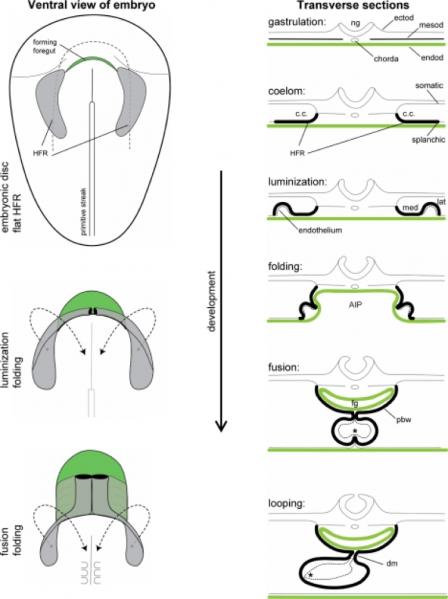
Figure 3
(ventral, transverse views)
Bilateral heart-forming region (shown in gray) swings toward ventral and medial to progressively fuse at the midline. - AIP anterior intestinal portal - c.c. coelomic cavity - dm dorsal mesocardium - ectod ectoderm - endod endoderm -fg foregut - HFR heart-forming region - lat lateral - med medial - mesod mesoderm - ng neural groove - pbw pericardial back wall, * contact between endocardium and myocardium
File history
Click on a date/time to view the file as it appeared at that time.
| Date/Time | Thumbnail | Dimensions | User | Comment | |
|---|---|---|---|---|---|
| current | 23:03, 17 September 2017 | | 772 × 1,032 (301 KB) | Z5076019 (talk | contribs) | [https://www.ncbi.nlm.nih.gov/pmc/articles/PMC2691808/pdf/246_2008_Article_9369.pdf] Received: 9 December 2008 / Accepted: 22 December 2008 / Published online: 30 January 2009 Ó The Author(s) 2009. This article is published with open access at Sprin... |
You cannot overwrite this file.
File usage
There are no pages that use this file.