File:Heart Tube Fusion Model of Early Development.png

Original file (772 × 1,032 pixels, file size: 301 KB, MIME type: image/png)
Description
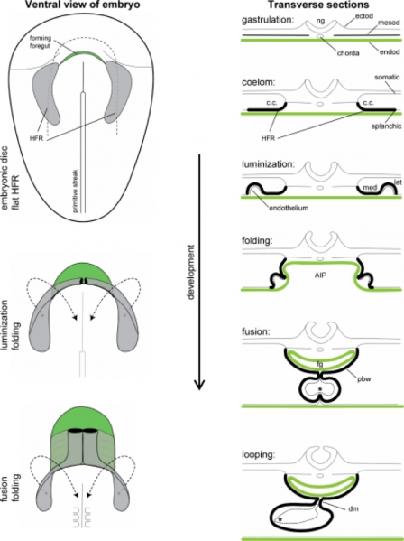
Figure 3
(ventral, transverse views)
Bilateral heart-forming region (shown in gray) swings toward ventral and medial to progressively fuse at the midline. - AIP anterior intestinal portal - c.c. coelomic cavity - dm dorsal mesocardium - ectod ectoderm - endod endoderm -fg foregut - HFR heart-forming region - lat lateral - med medial - mesod mesoderm - ng neural groove - pbw pericardial back wall, * contact between endocardium and myocardium
References
<pubmed> PMC2691808 </pubmed>
Copyright
Received: 9 December 2008 / Accepted: 22 December 2008 / Published online: 30 January 2009 Ó The Author(s) 2009. This article is published with open access at Springerlink.com
- Note - This image was originally uploaded as part of an undergraduate science student project and may contain inaccuracies in either description or acknowledgements. Students have been advised in writing concerning the reuse of content and may accidentally have misunderstood the original terms of use. If image reuse on this non-commercial educational site infringes your existing copyright, please contact the site editor for immediate removal.
File history
Click on a date/time to view the file as it appeared at that time.
| Date/Time | Thumbnail | Dimensions | User | Comment | |
|---|---|---|---|---|---|
| current | 23:03, 17 September 2017 | | 772 × 1,032 (301 KB) | Z5076019 (talk | contribs) | [https://www.ncbi.nlm.nih.gov/pmc/articles/PMC2691808/pdf/246_2008_Article_9369.pdf] Received: 9 December 2008 / Accepted: 22 December 2008 / Published online: 30 January 2009 Ó The Author(s) 2009. This article is published with open access at Sprin... |
You cannot overwrite this file.
File usage
There are no pages that use this file.