File:Heart Tube Fusion.jpg
From Embryology
Size of this preview: 800 × 587 pixels. Other resolution: 1,551 × 1,139 pixels.
Original file (1,551 × 1,139 pixels, file size: 125 KB, MIME type: image/jpeg)
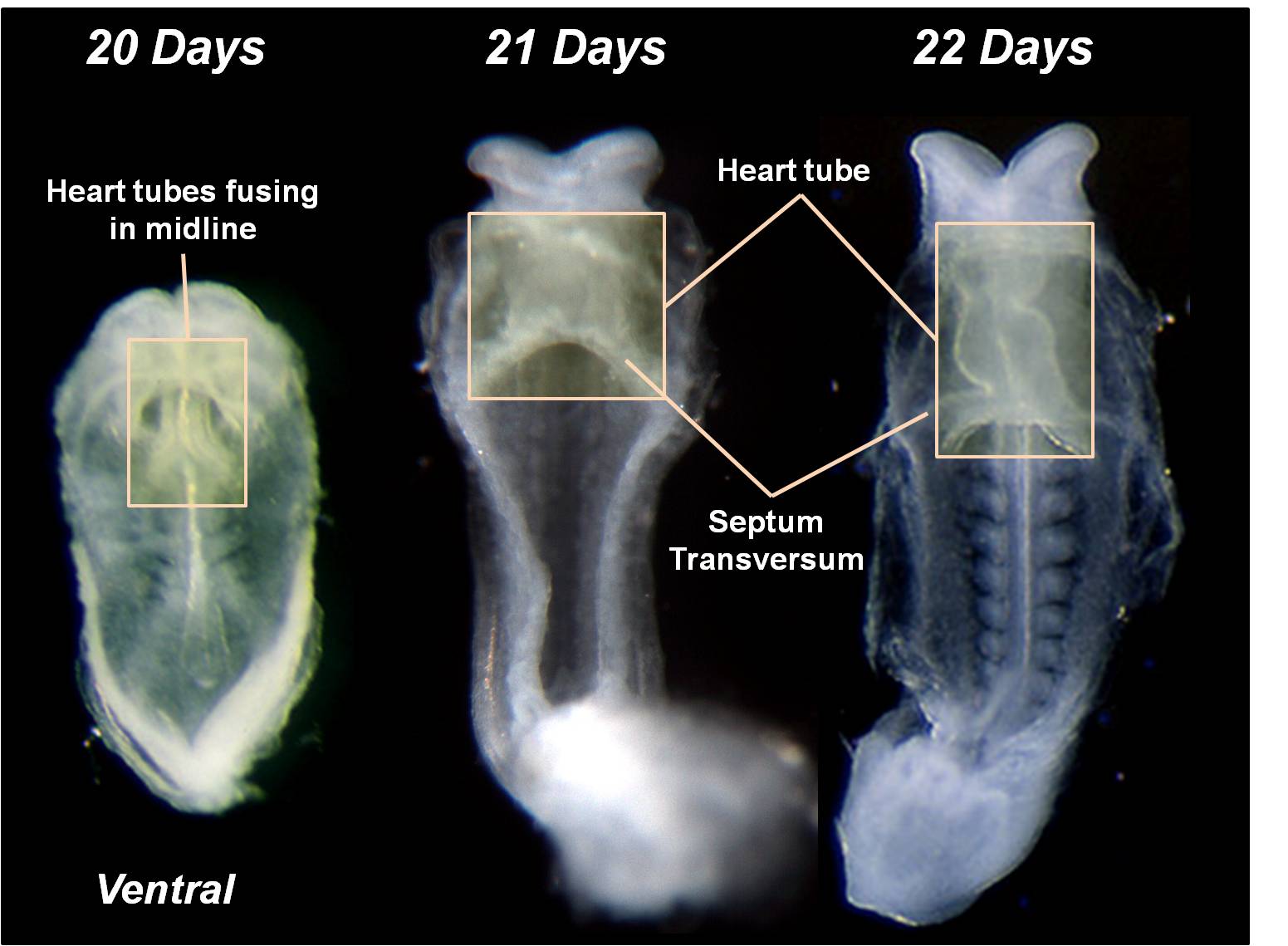
Image Source: Scanning electron micrographs of the Carnegie stages of the early human embryos are reproduced with the permission of Prof Kathy Sulik, from embryos collected by Dr. Vekemans and Tania Attié-Bitach. Images are for educational purposes only and cannot be reproduced electronically or in writing without permission.
The primordial heart tubes fuse in the midline to form a single ventral heart tube. Fusion begins cranially and extends caudally.
File history
Click on a date/time to view the file as it appeared at that time.
| Date/Time | Thumbnail | Dimensions | User | Comment | |
|---|---|---|---|---|---|
| current | 10:41, 14 March 2010 | | 1,551 × 1,139 (125 KB) | Z3212774 (talk | contribs) | category:Heart ILP {{Template:SEM}} The primordial heart tubes fuse in the midline to form a single ventral heart tube. Fusion begins cranially and extends caudally. |
You cannot overwrite this file.
File usage
The following 15 pages use this file:
- 2009 Lecture 21
- 2010 BGD Lecture - Development of the Embryo/Fetus 1
- 2010 Lecture 21
- ANAT2341 Lab 4 - Early Cardiovascular Development
- BGDA Lecture - Development of the Embryo/Fetus 1
- BGDA Practical 7 - Week 4
- Cardiovascular - Arterial Development
- Cardiovascular - Venous Development
- Cardiovascular System Development
- Fetal ECHO Meeting 2012
- Human Embryo SEM
- Human System Development
- Intermediate - Primordial Heart Tube
- Lecture - Week 3 Development
- RPAH Cardiac Embryology 2014