File:GnRH receptors (GnRHRs) and spatial expression patterns of gnrhr genes.png: Difference between revisions
(http://www.plosone.org/article/info%3Adoi%2F10.1371%2Fjournal.pone.0041955) |
No edit summary |
||
| Line 1: | Line 1: | ||
http://www.plosone.org/article/info%3Adoi%2F10.1371%2Fjournal.pone.0041955 | http://www.plosone.org/article/info%3Adoi%2F10.1371%2Fjournal.pone.0041955 | ||
Figure 1. Expression of gnrh genes in the Ciona intestinalis larvae. | |||
Spatial expression patterns of Ci-gnrh1 (A, C, and D) and Ci-gnrh2 (B, E, G, and H) in a whole body (A, B, and G) or trunk region (C-F, and H) of C. intestinalis larvae. The gene expression was visualized by whole-mount in situ hybridization (A, B, C, and E) or by GFP reporter expression under the control of the regulatory region of Ci-gnrh1 (D, G, and H). (F) An unstained control larva showing the otolith (ot) and ocellus (oc) pigment cells in the brain vesicle. White arrowheads in (D) indicate GFP-positive axons of papillar neurons (pn). The white arrowhead in (H) indicates a GFP positive neurite. bv, brain vesicle; nc, nerve cord; rten, rostoral trunk epidermal neuron; mg, motor ganglion. Scale bars: 100 µm in (A); 50 µm in (C). | |||
==Reference== | |||
Citation: Kusakabe TG, Sakai T, Aoyama M, Kitajima Y, Miyamoto Y, et al. (2012) A Conserved Non-Reproductive GnRH System in Chordates. PLoS ONE 7(7): e41955. doi:10.1371/journal.pone.0041955 | |||
Copyright: © 2012 Kusakabe et al. This is an open-access article distributed under the terms of the Creative Commons Attribution License, which permits unrestricted use, distribution, and reproduction in any medium, provided the original author and source are credited. | |||
_and_spatial_expression_patterns_of_gnrhr_genes.png&oldid=96992)
_and_spatial_expression_patterns_of_gnrhr_genes.png&action=edit&oldid=96992)
_and_spatial_expression_patterns_of_gnrhr_genes.png&oldid=97000)
_and_spatial_expression_patterns_of_gnrhr_genes.png&action=edit&oldid=97000)
_and_spatial_expression_patterns_of_gnrhr_genes.png&diff=next&oldid=97000)
Revision as of 12:01, 1 August 2012
http://www.plosone.org/article/info%3Adoi%2F10.1371%2Fjournal.pone.0041955
Figure 1. Expression of gnrh genes in the Ciona intestinalis larvae.
Spatial expression patterns of Ci-gnrh1 (A, C, and D) and Ci-gnrh2 (B, E, G, and H) in a whole body (A, B, and G) or trunk region (C-F, and H) of C. intestinalis larvae. The gene expression was visualized by whole-mount in situ hybridization (A, B, C, and E) or by GFP reporter expression under the control of the regulatory region of Ci-gnrh1 (D, G, and H). (F) An unstained control larva showing the otolith (ot) and ocellus (oc) pigment cells in the brain vesicle. White arrowheads in (D) indicate GFP-positive axons of papillar neurons (pn). The white arrowhead in (H) indicates a GFP positive neurite. bv, brain vesicle; nc, nerve cord; rten, rostoral trunk epidermal neuron; mg, motor ganglion. Scale bars: 100 µm in (A); 50 µm in (C).
Reference
Citation: Kusakabe TG, Sakai T, Aoyama M, Kitajima Y, Miyamoto Y, et al. (2012) A Conserved Non-Reproductive GnRH System in Chordates. PLoS ONE 7(7): e41955. doi:10.1371/journal.pone.0041955
Copyright: © 2012 Kusakabe et al. This is an open-access article distributed under the terms of the Creative Commons Attribution License, which permits unrestricted use, distribution, and reproduction in any medium, provided the original author and source are credited.
File history
Click on a date/time to view the file as it appeared at that time.
| Date/Time | Thumbnail | Dimensions | User | Comment | |
|---|---|---|---|---|---|
| current | 11:59, 1 August 2012 | 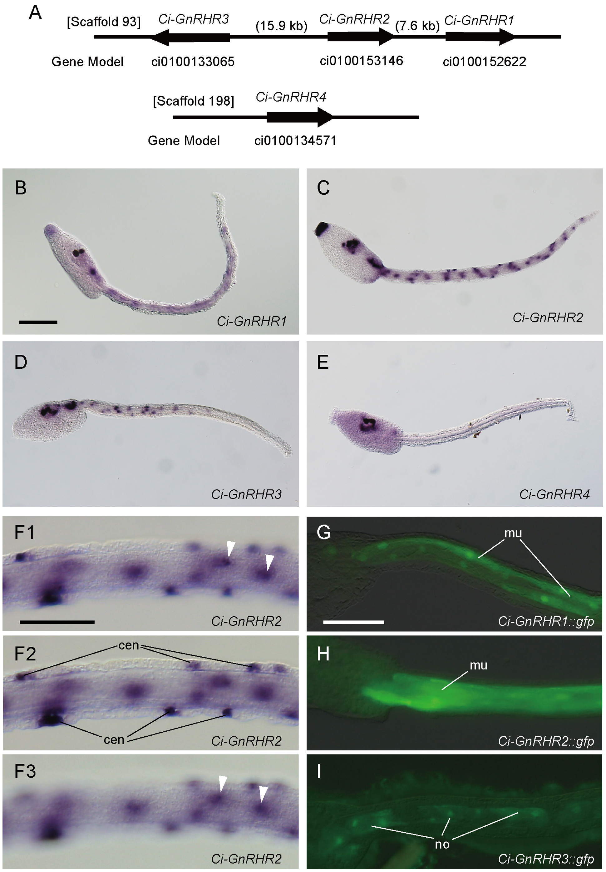 | 1,941 × 2,790 (6.16 MB) | Z3333794 (talk | contribs) | http://www.plosone.org/article/info%3Adoi%2F10.1371%2Fjournal.pone.0041955 |
You cannot overwrite this file.
File usage
The following page uses this file:
_and_spatial_expression_patterns_of_gnrhr_genes.png&oldid=97000)