File:Formation of the lens 2.jpg
From Embryology
Size of this preview: 800 × 450 pixels. Other resolution: 1,152 × 648 pixels.
Original file (1,152 × 648 pixels, file size: 94 KB, MIME type: image/jpeg)
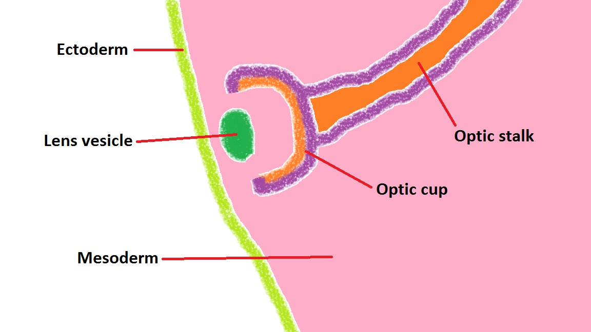
A diagram showing completed invagination of the thickened lens placode of the ectoderm into the mesoderm, forming the lens vesicle. The combined structure of the optic cup and lens vesicle can now be referred to as the optic globe.
Copyright: This is a student drawn image and is free for non-profit reuse.
- Note - This image was originally uploaded as part of an undergraduate science student project and may contain inaccuracies in either description or acknowledgements. Students have been advised in writing concerning the reuse of content and may accidentally have misunderstood the original terms of use. If image reuse on this non-commercial educational site infringes your existing copyright, please contact the site editor for immediate removal.
File history
Click on a date/time to view the file as it appeared at that time.
| Date/Time | Thumbnail | Dimensions | User | Comment | |
|---|---|---|---|---|---|
| current | 17:17, 27 September 2012 | | 1,152 × 648 (94 KB) | Z3373894 (talk | contribs) | A diagram showing completed invagination of the thickened lens placode of the ectoderm into the mesoderm, forming the lens vesicle. The combined structure of the optic cup and lens vesicle can now be referred to as the optic globe. Copyright: This is a s |
You cannot overwrite this file.
File usage
The following 2 pages use this file: