File:Effect-of-vegf-on-retinal-vasculature.JPG
Effect-of-vegf-on-retinal-vasculature.JPG (374 × 600 pixels, file size: 50 KB, MIME type: image/jpeg)
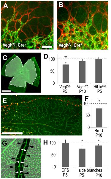
Effects of astrocyte-derived VEGF on retinal vascular development
"Immunohistochemistry, visualizing endothelial cells (claudin 5, green in A, B, C, E, G) and retinal astrocytes (GFAP, red in A, B), shows that angiogenic sprouting at the leading edge of the growing vascular plexus appears normal in control (A) and mutant (B) animals lacking astrocyte derived VEGF. (C, D) Measurement of the surface area of the retinal vasculature (C) showed significantly reduced spreading in Vegfc/c mutants at P5 but not at P10 and not in P5 Hif1ac/c mutants at P5. (E) AT P10 proliferation (anti-BrdU in red) occurs predominantly in veins and is reduced in animals lacking astrocyte-derived VEGF in comparison to littermate controls. (G–H) Endothelial cell survival near arteries was assessed by measuring the width of the capillary free zone (CFS, white arrows) and the number of artery side branches (black arrowheads). (H) In mutant animals CFS width was not affected but the number of side branches was reduced. Scale bars are 50µm in A, 1000µm in C and 200µm in G; * is p<0.05 and ** is p<0.01."
doi:10.1371/journal.pone.0011863.g002
Reference
<pubmed>20686684</pubmed>
Copyright
© 2010 Scott et al. This is an open-access article distributed under the terms of the Creative Commons Attribution License, which permits unrestricted use, distribution, and reproduction in any medium, provided the original author and source are credited.
- Note - This image was originally uploaded as part of an undergraduate science student project and may contain inaccuracies in either description or acknowledgements. Students have been advised in writing concerning the reuse of content and may accidentally have misunderstood the original terms of use. If image reuse on this non-commercial educational site infringes your existing copyright, please contact the site editor for immediate removal.
File history
Click on a date/time to view the file as it appeared at that time.
| Date/Time | Thumbnail | Dimensions | User | Comment | |
|---|---|---|---|---|---|
| current | 06:01, 3 October 2012 | | 374 × 600 (50 KB) | Z3370664 (talk | contribs) | "Effects of astrocyte-derived VEGF on retinal vascular development." "Immunohistochemistry, visualizing endothelial cells (claudin 5, green in A, B, C, E, G) and retinal astrocytes (GFAP, red in A, B), shows that angiogenic sprouting at the leading edge |
You cannot overwrite this file.
File usage
The following page uses this file: