File:Differentiation of NCC.jpg
From Embryology
Size of this preview: 800 × 373 pixels. Other resolution: 1,168 × 545 pixels.
Original file (1,168 × 545 pixels, file size: 75 KB, MIME type: image/jpeg)
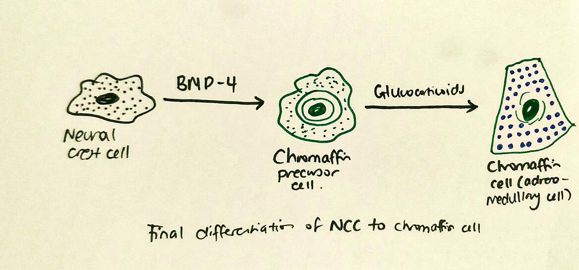
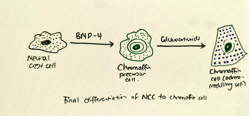
The neural crest cells become chromaffin precursor cells under exposure to BMP-4, and their growth and postnatal health is maintained by the secretion of glucocorticoids.
This image was inspired by the following figure on the link below, however the information was altered slightly, due to recent studies showing that glucocorticoids do not initially cause neural crest cells to become chromaffin cells, but rather BMP-4 protein[1]:
https://www.ncbi.nlm.nih.gov/books/NBK10065/figure/A3132/?report=objectonly [2].
- ↑ Unsicker K, Huber K, Schober A & Kalcheim C. (2013). Resolved and open issues in chromaffin cell development. Mech. Dev. , 130, 324-9. PMID: 23220335 DOI.
- ↑ Gilbert SF. Developmental Biology. 6th edition. Sunderland (MA): Sinauer Associates; 2000. The Neural Crest. Available from: https://www.ncbi.nlm.nih.gov/books/NBK10065/
File history
Click on a date/time to view the file as it appeared at that time.
| Date/Time | Thumbnail | Dimensions | User | Comment | |
|---|---|---|---|---|---|
| current | 17:31, 10 October 2018 | | 1,168 × 545 (75 KB) | Z5091101 (talk | contribs) | The neural crest cells become chromaffin precursor cells under exposure to BMP-4, and their growth and postnatal health is maintained by the secretion of glucocorticoids. |
You cannot overwrite this file.
File usage
The following 2 pages use this file: