File:Devt of external ear.JPG: Difference between revisions
From Embryology
No edit summary |
No edit summary |
||
| Line 11: | Line 11: | ||
<pubmed>17104502</pubmed> | <pubmed>17104502</pubmed> | ||
{{Template:Student Image}} | |||
Revision as of 16:42, 28 September 2012
The development of Pinna
The image illustrates the development of pinna.
1 tragus 2 helix 3 cymba concha 4 concha 5 antihelix 6 antitragus
<pubmed>17104502</pubmed>
- Note - This image was originally uploaded as part of an undergraduate science student project and may contain inaccuracies in either description or acknowledgements. Students have been advised in writing concerning the reuse of content and may accidentally have misunderstood the original terms of use. If image reuse on this non-commercial educational site infringes your existing copyright, please contact the site editor for immediate removal.
File history
Click on a date/time to view the file as it appeared at that time.
| Date/Time | Thumbnail | Dimensions | User | Comment | |
|---|---|---|---|---|---|
| current | 11:42, 26 September 2012 | 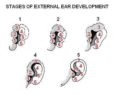 | 399 × 320 (20 KB) | Z3333794 (talk | contribs) |
You cannot overwrite this file.
File usage
The following 2 pages use this file: