File:Detection and Localisation of HPV in Sperms.png
Detection_and_Localisation_of_HPV_in_Sperms.png (600 × 238 pixels, file size: 288 KB, MIME type: image/png)
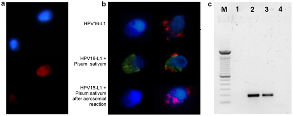
Detection and Localisation of Human Papilloma Virus (HPV) in Spermatozoa
a. Fluorescence in situ hybridization (fluorescence microscope) for HPV DNA on sperm from a patient with HPV16 in semen. Infected and noninfected sperm are shown. Red: HPV DNA (Texas red); blue: nuclear staining (DAPI).
b. Immunofluorescence (confocal fluorescence microscope) for HPV16 capsid protein L1 on sperm from a control (left) and a patient with HPV16 in semen (right). Upper panel, L1 antibody; central panel, L1 antibody and Pisum Sativum (acrosome); lower panel, L1 antibody and Pisum Sativum after induction of the acrosome reaction. Red: HPV16 L1; green: Pisum Sativum; blue: nuclear staining (DAPI).
c. PCR for HPV E7 gene from sperm DNA. Lane M: DNA marker (100 bp); 1: negative control (no template); 2: positive control (sperm transfected with recombinant plasmid pIRES2-AcGFP1-E6E7); 3: sperm from a patient with HPV16 in semen; 4: sperm from a control subject.
Reference
<pubmed>21408100</pubmed>
Copyright
© 2011 Carlo et al. This is an open-access article distributed under the terms of the Creative Commons Attribution License, which permits unrestricted use, distribution, and reproduction in any medium, provided the original author and source are credited.
- Note - This image was originally uploaded as part of an undergraduate science student project and may contain inaccuracies in either description or acknowledgements. Students have been advised in writing concerning the reuse of content and may accidentally have misunderstood the original terms of use. If image reuse on this non-commercial educational site infringes your existing copyright, please contact the site editor for immediate removal.
Cite this page: Hill, M.A. (2024, April 27) Embryology Detection and Localisation of HPV in Sperms.png. Retrieved from https://embryology.med.unsw.edu.au/embryology/index.php/File:Detection_and_Localisation_of_HPV_in_Sperms.png
- © Dr Mark Hill 2024, UNSW Embryology ISBN: 978 0 7334 2609 4 - UNSW CRICOS Provider Code No. 00098G
File history
Click on a date/time to view the file as it appeared at that time.
| Date/Time | Thumbnail | Dimensions | User | Comment | |
|---|---|---|---|---|---|
| current | 15:28, 5 August 2012 | 600 × 238 (288 KB) | Z3332863 (talk | contribs) | Detection and localization of HPV in human sperm. a. Fluorescence in situ hybridization (fluorescence microscope) for HPV DNA on sperm from a patient with HPV16 in semen. Infected and noninfected sperm are shown. Red: HPV DNA (Texas red); blue: nuclear s |
You cannot overwrite this file.
File usage
The following page uses this file: