File:Auditory Cortex Location - Comparison Between Control Subject and WS Subject.jpg: Difference between revisions
(Original File Name: journal.pone.0012326.g002.jpg Legend: Auditory Cortex Location - Comparison Between Control Subject and WS Subject Citation: Wengenroth M, Blatow M, Bendszus M, Schneider P (2010) Leftward Laterlization of Auditory Cortex Underlies H) |
No edit summary |
||
| Line 1: | Line 1: | ||
==Auditory Cortex Location - Comparison Between Control Subject and WS Subject== | |||
Original File Name: journal.pone.0012326.g002.jpg | Original File Name: journal.pone.0012326.g002.jpg | ||
Legend: Auditory Cortex Location - Comparison Between Control Subject and WS Subject | Legend: Auditory Cortex Location - Comparison Between Control Subject and WS Subject | ||
Original Legend: Figure 2. Anatomical landmarks of the auditory cortex. | Original Legend: Figure 2. Anatomical landmarks of the auditory cortex. | ||
Auditory cortex (AC) of one control person (a–c) and one WS subject (d–e). Sagittal MR image at TAL x = 50 (a,d: left side of the image is the anterior part of the brain). Segmented STG (b,e) including Heschl's gyrus (HG; marked orange), planum temporale (PT; marked yellow) and two posterior duplications of HG in the WS subject (D; marked green). Three-dimensional surface reconstruction of right AC (c,f) reveals anatomical features and individual peculiarities such as D (f) or medial Heschl's sulcus (mHS; c). FTS = first transverse sulcus; HS = Heschl's sulcus; aSTG = anterior superior temporal gyrus. | Auditory cortex (AC) of one control person (a–c) and one WS subject (d–e). Sagittal MR image at TAL x = 50 (a,d: left side of the image is the anterior part of the brain). Segmented STG (b,e) including Heschl's gyrus (HG; marked orange), planum temporale (PT; marked yellow) and two posterior duplications of HG in the WS subject (D; marked green). Three-dimensional surface reconstruction of right AC (c,f) reveals anatomical features and individual peculiarities such as D (f) or medial Heschl's sulcus (mHS; c). FTS = first transverse sulcus; HS = Heschl's sulcus; aSTG = anterior superior temporal gyrus. | ||
===Reference=== | |||
Citation: Wengenroth M, Blatow M, Bendszus M, Schneider P (2010) Leftward Laterlization of Auditory Cortex Underlies Holistic Sound Perception in Williams Syndrome. PLoS ONE 5(8): e12326. doi:10.1371/journal.pone.0012326 | |||
PMID:20808792 | |||
Copyright: © 2010 Wengenroth et al. This is an open-access article distributed under the terms of the Creative Commons Attribution License, which permits unrestricted use, distribution, and reproduction in any medium, provided the original author and source are credited. | Copyright: © 2010 Wengenroth et al. This is an open-access article distributed under the terms of the Creative Commons Attribution License, which permits unrestricted use, distribution, and reproduction in any medium, provided the original author and source are credited. | ||
Revision as of 15:33, 17 September 2011
Auditory Cortex Location - Comparison Between Control Subject and WS Subject
Original File Name: journal.pone.0012326.g002.jpg
Legend: Auditory Cortex Location - Comparison Between Control Subject and WS Subject
Original Legend: Figure 2. Anatomical landmarks of the auditory cortex.
Auditory cortex (AC) of one control person (a–c) and one WS subject (d–e). Sagittal MR image at TAL x = 50 (a,d: left side of the image is the anterior part of the brain). Segmented STG (b,e) including Heschl's gyrus (HG; marked orange), planum temporale (PT; marked yellow) and two posterior duplications of HG in the WS subject (D; marked green). Three-dimensional surface reconstruction of right AC (c,f) reveals anatomical features and individual peculiarities such as D (f) or medial Heschl's sulcus (mHS; c). FTS = first transverse sulcus; HS = Heschl's sulcus; aSTG = anterior superior temporal gyrus.
Reference
Citation: Wengenroth M, Blatow M, Bendszus M, Schneider P (2010) Leftward Laterlization of Auditory Cortex Underlies Holistic Sound Perception in Williams Syndrome. PLoS ONE 5(8): e12326. doi:10.1371/journal.pone.0012326
PMID:20808792
Copyright: © 2010 Wengenroth et al. This is an open-access article distributed under the terms of the Creative Commons Attribution License, which permits unrestricted use, distribution, and reproduction in any medium, provided the original author and source are credited.
- Note - This image was originally uploaded as part of an undergraduate science student project and may contain inaccuracies in either description or acknowledgements. Students have been advised in writing concerning the reuse of content and may accidentally have misunderstood the original terms of use. If image reuse on this non-commercial educational site infringes your existing copyright, please contact the site editor for immediate removal.
File history
Click on a date/time to view the file as it appeared at that time.
| Date/Time | Thumbnail | Dimensions | User | Comment | |
|---|---|---|---|---|---|
| current | 17:24, 16 August 2011 | 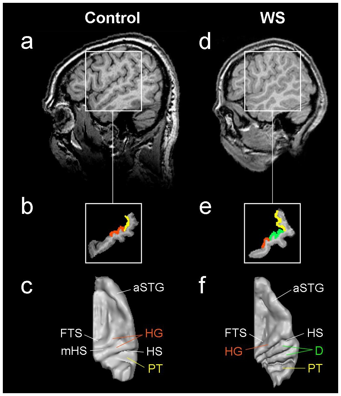 | 1,084 × 1,261 (234 KB) | Z3331469 (talk | contribs) | Original File Name: journal.pone.0012326.g002.jpg Legend: Auditory Cortex Location - Comparison Between Control Subject and WS Subject Citation: Wengenroth M, Blatow M, Bendszus M, Schneider P (2010) Leftward Laterlization of Auditory Cortex Underlies H |
You cannot overwrite this file.
File usage
The following 3 pages use this file: