File:Astrocyte-vegf-deletion.JPG
Astrocyte-vegf-deletion.JPG (495 × 600 pixels, file size: 75 KB, MIME type: image/jpeg)
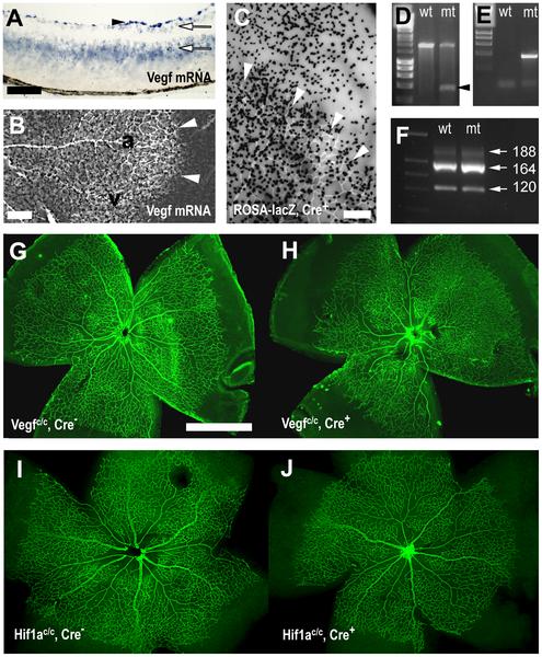
"Astrocyte specific deletion of VEGF."
"(A, B) In situ hybridisation showed Vegf mRNA expression in a retinal cross-section (A) and a retinal whole mount (B) at P5. (A) Vegf mRNA was most strongly expressed in retinal astrocytes (arrowhead), and weakly in retinal ganglion cells and the inner nuclear layer (arrows). (B) Retinal astrocytes expressed VEGF strongest distally to the edge of growing vascular plexus (white arrowheads), at intermediate levels around veins (v) and weakest along arteries (a). (C) Transgenic mice expressing Cre recombinase in retinal astrocytes (Gfap-Cre mice) showed in a lacZ reporter strain recombination activity (black X-gal stain in C) in retinal astrocytes proximally and distally to the growing edge of the vascular plexus (arrowheads in C). (D, E) Cre recombination in Vegfc/c mice, crossed with Gfap-Cre mice, was demonstrated by PCR across the deleted region in genomic DNA from retinas in Cre positive (mt) and Cre negative (wt) animals (D). PCR on tail DNA identified Cre negative and positive animals (E). (F) PCR on cDNA from retinal mRNA showed unchanged VEGF isoform ratios in control and mutant animals. Astrocyte deletion of VEGF led to subtle abnormalities in the P5 retinal vasculature (stained with an anti-claudin 5 antibody G–J) in Cre positive animals (H) compared to wild type litter mates (G). In contrast, deletion of HIF1α in retinal astrocytes caused no such abnormalities (I–J). Scale bars are 100µm in A, 200µm in B, C and 1000µm in G."
doi:10.1371/journal.pone.0011863.g001
Source: <pubmed>20686684</pubmed>
Copyright: © 2010 Scott et al. This is an open-access article distributed under the terms of the Creative Commons Attribution License, which permits unrestricted use, distribution, and reproduction in any medium, provided the original author and source are credited.
- Note - This image was originally uploaded as part of an undergraduate science student project and may contain inaccuracies in either description or acknowledgements. Students have been advised in writing concerning the reuse of content and may accidentally have misunderstood the original terms of use. If image reuse on this non-commercial educational site infringes your existing copyright, please contact the site editor for immediate removal.
File history
Click on a date/time to view the file as it appeared at that time.
| Date/Time | Thumbnail | Dimensions | User | Comment | |
|---|---|---|---|---|---|
| current | 06:00, 3 October 2012 | | 495 × 600 (75 KB) | Z3370664 (talk | contribs) | "Astrocyte specific deletion of VEGF." "(A, B) In situ hybridisation showed Vegf mRNA expression in a retinal cross-section (A) and a retinal whole mount (B) at P5. (A) Vegf mRNA was most strongly expressed in retinal astrocytes (arrowhead), and weakly i |
You cannot overwrite this file.
File usage
The following page uses this file: