File:Anatomical diagram of testes.jpg: Difference between revisions
From Embryology
(Z3417753 uploaded a new version of "File:Anatomical diagram of testes.jpg") |
(Z3417753 uploaded a new version of "File:Anatomical diagram of testes.jpg") |
(No difference)
| |
Revision as of 20:29, 20 October 2014
Student drawn image of the testes, epididymis, ductus deferens and spermatic cord.
File history
Click on a date/time to view the file as it appeared at that time.
| Date/Time | Thumbnail | Dimensions | User | Comment | |
|---|---|---|---|---|---|
| current | 20:29, 20 October 2014 |  | 647 × 834 (237 KB) | Z3417753 (talk | contribs) | Reverted to version as of 10:27, 20 October 2014 |
| 20:28, 20 October 2014 |  | 647 × 834 (237 KB) | Z3417753 (talk | contribs) | Reverted to version as of 10:08, 20 October 2014 | |
| 20:27, 20 October 2014 |  | 647 × 834 (237 KB) | Z3417753 (talk | contribs) | Reverted to version as of 10:08, 20 October 2014 | |
| 20:08, 20 October 2014 |  | 647 × 834 (235 KB) | Z3417753 (talk | contribs) | Reverted to version as of 10:05, 20 October 2014 | |
| 20:08, 20 October 2014 | 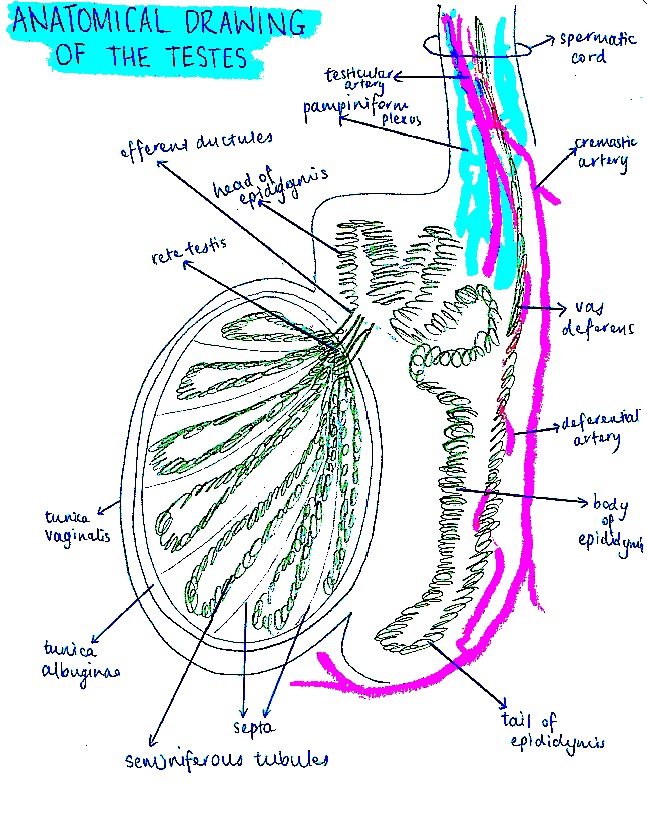 | 647 × 834 (237 KB) | Z3417753 (talk | contribs) | ||
| 20:05, 20 October 2014 | 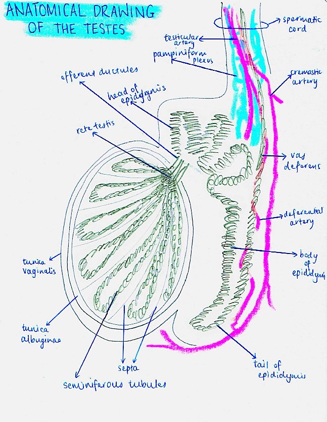 | 647 × 834 (235 KB) | Z3417753 (talk | contribs) | ||
| 19:24, 20 October 2014 |  | 647 × 834 (472 KB) | Z3417753 (talk | contribs) | Student drawn image of the testes, epididymis, ductus deferens and spermatic cord. |
You cannot overwrite this file.
File usage
The following 2 pages use this file: