File:Amnion fold development in chicken embryos.jpg
Amnion_fold_development_in_chicken_embryos.jpg (786 × 443 pixels, file size: 94 KB, MIME type: image/jpeg)
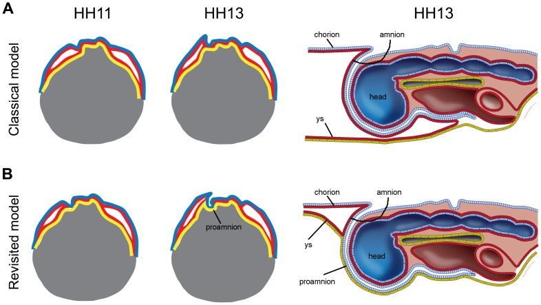
Two models (classical and revisited) for anterior amnion fold development in chicken embryos
(A) Sagittal scheme, representing the classical model, of HH11 and HH13 chicken embryos adapted from a Developmental Biology textbook [36] and a detail view of the head region at HH13. (B) Sagittal scheme, representing the revisited model, of HH11 and HH13 chicken embryos adapted from Shore and Pickering [9]. In both schemes, ectoderm is blue, mesoderm is red, endoderm is yellow and yolk is gray. Abbreviations: ys, yolk sac.
http://dx.doi.org/10.1371/journal.pone.0092672
Reference
<pubmed>24647352</pubmed>
De Melo Bernardo A, Chuva de Sousa Lopes SM (2014) The Involvement of the Proamnion in the Development of the Anterior Amnion Fold in the Chicken. PLoS ONE 9(3): e92672. doi:10.1371/journal.pone.0092672
Copyright
© 2014 de Melo Bernardo, Chuva de Sousa Lopes. This is an open-access article distributed under the terms of the Creative Commons Attribution License, which permits unrestricted use, distribution, and reproduction in any medium, provided the original author and source are credited.
- Note - This image was originally uploaded as part of an undergraduate science student project and may contain inaccuracies in either description or acknowledgements. Students have been advised in writing concerning the reuse of content and may accidentally have misunderstood the original terms of use. If image reuse on this non-commercial educational site infringes your existing copyright, please contact the site editor for immediate removal.
File history
Click on a date/time to view the file as it appeared at that time.
| Date/Time | Thumbnail | Dimensions | User | Comment | |
|---|---|---|---|---|---|
| current | 22:05, 14 August 2016 | | 786 × 443 (94 KB) | Z5020373 (talk | contribs) | PMID 24647352 (A) Sagittal scheme, representing the classical model, of HH11 and HH13 chicken embryos adapted from a Developmental Biology textbook [36] and a detail view of the head region at HH13. (B) Sagittal scheme, representing the revisited mod... |
You cannot overwrite this file.
File usage
The following page uses this file: