File:8cellembryo.jpg
From Embryology
Size of this preview: 391 × 599 pixels. Other resolution: 424 × 650 pixels.
Original file (424 × 650 pixels, file size: 42 KB, MIME type: image/jpeg)
Citation: Viebahn, C. (n.d.) Formation of the body axes in the early mammalian embryo. Anatomy & Embryology of Universität Göttingenm, Göttingenm.
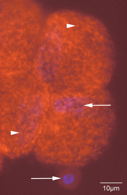
Immunofluroescence staining (red) of the mitochondrial antigen PG2 outlining individual mitochondria (arrowheads) in 4 blastomeres of a 8-cell rabbit embryo. White arrows mark DAPI-stained DNA in nuclei of blastomere (top) and polar body (bottom).
File history
Click on a date/time to view the file as it appeared at that time.
| Date/Time | Thumbnail | Dimensions | User | Comment | |
|---|---|---|---|---|---|
| current | 16:46, 14 September 2009 | | 424 × 650 (42 KB) | Z3186093 (talk | contribs) | [http://www.embryologie.uni-goettingen.de/data/cviebah/PG2.jpg/ Image Location] Citation: Viebahn, C. (n.d.) Formation of the body axes in the early mammalian embryo. Anatomy & Embryology of Universität Göttingenm, Göttingenm. [http://www.embryolo |
You cannot overwrite this file.
File usage
The following page uses this file: