File:3–11 SS caudal foregut endoderm.png

Original file (1,347 × 1,855 pixels, file size: 315 KB, MIME type: image/png)
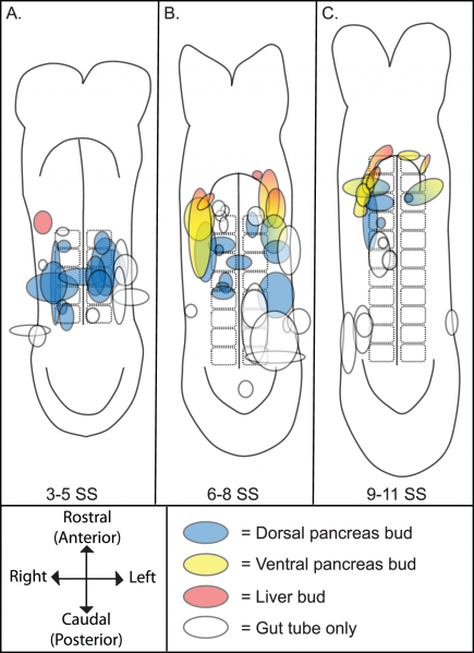
3–11 SS caudal foregut endoderm
DiI labelling of the endoderm of individual 3-11 SS embryos and culturing at approx 9.5 dpc. Position and size of the DiI labelled endoderm from a single embryo is illustrated through its shape. The colour indicates which organ bud contributed to the end of culture. Simple compass is provided indicating the embryonic axis.
Reference
<pubmed>22815796</pubmed>
Copyright: © 2012 Angelo et al. This is an open-access article distributed under the terms of the Creative Commons Attribution License, which permits unrestricted use, distribution, and reproduction in any medium, provided the original author and source are credited.
- Note - This image was originally uploaded as part of an undergraduate science student project and may contain inaccuracies in either description or acknowledgements. Students have been advised in writing concerning the reuse of content and may accidentally have misunderstood the original terms of use. If image reuse on this non-commercial educational site infringes your existing copyright, please contact the site editor for immediate removal.
File history
Click on a date/time to view the file as it appeared at that time.
| Date/Time | Thumbnail | Dimensions | User | Comment | |
|---|---|---|---|---|---|
| current | 23:53, 23 October 2014 | | 1,347 × 1,855 (315 KB) | Z3414515 (talk | contribs) | ==3–11 SS caudal foregut endoderm== DiI labelling of the endoderm of individual 3-11 SS embryos and culturing at approx 9.5 dpc. Position and size of the DiI labelled endoderm from a single embryo is illustrated through its shape. The colour indicat... |
You cannot overwrite this file.
File usage
The following 2 pages use this file: