File:Spina BifidaMyelomeningocele.jpg
From Embryology
Size of this preview: 800 × 270 pixels. Other resolution: 886 × 299 pixels.
Original file (886 × 299 pixels, file size: 39 KB, MIME type: image/jpeg)
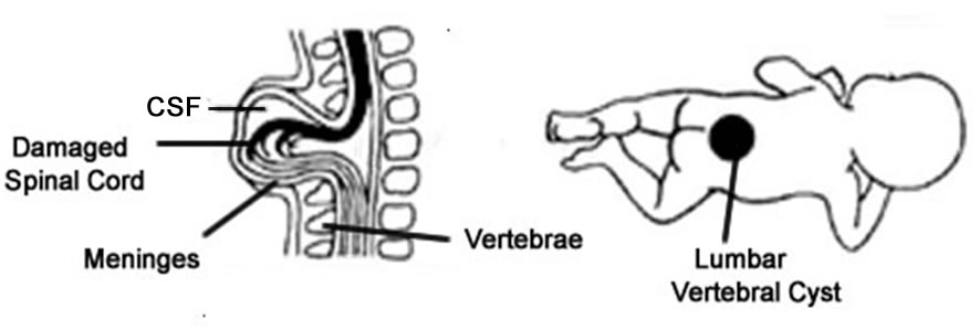
Modified image showing Spina Bifida Myelomeningocele characterized by split outer vertebrae with spinal cord and its meninges protruding from the divided vertebrae as a cyst. Commonly found at lumbar vertebral level
Original article
see Spina Bifida Meningocele image
File history
Yi efo/eka'e gwa ebo wo le nyangagi wuncin ye kamina wunga tinya nan
| Gwalagizhi | Nyangagi | Dimensions | User | Comment | |
|---|---|---|---|---|---|
| current | 14:29, 18 September 2009 | | 886 × 299 (39 KB) | Z3187802 (talk | contribs) | Reverted to version as of 03:28, 18 September 2009 |
| 14:29, 18 September 2009 | 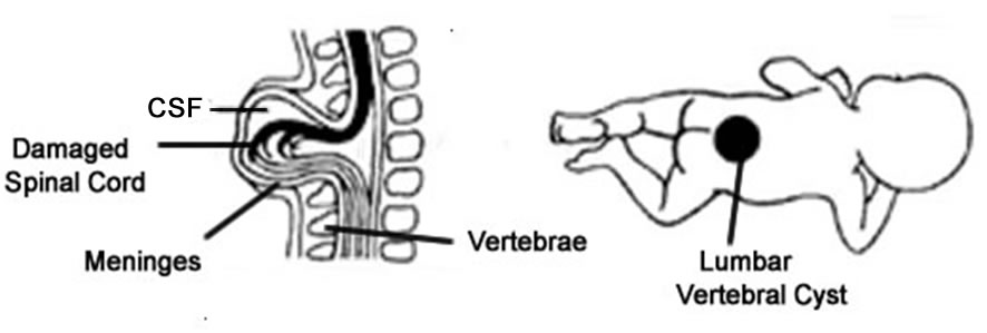 | 886 × 299 (39 KB) | Z3187802 (talk | contribs) | ||
| 14:28, 18 September 2009 | 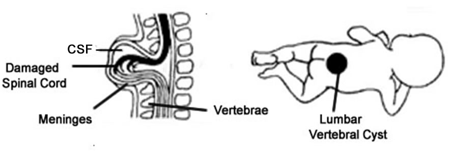 | 886 × 299 (39 KB) | Z3187802 (talk | contribs) | ||
| 06:04, 17 September 2009 | 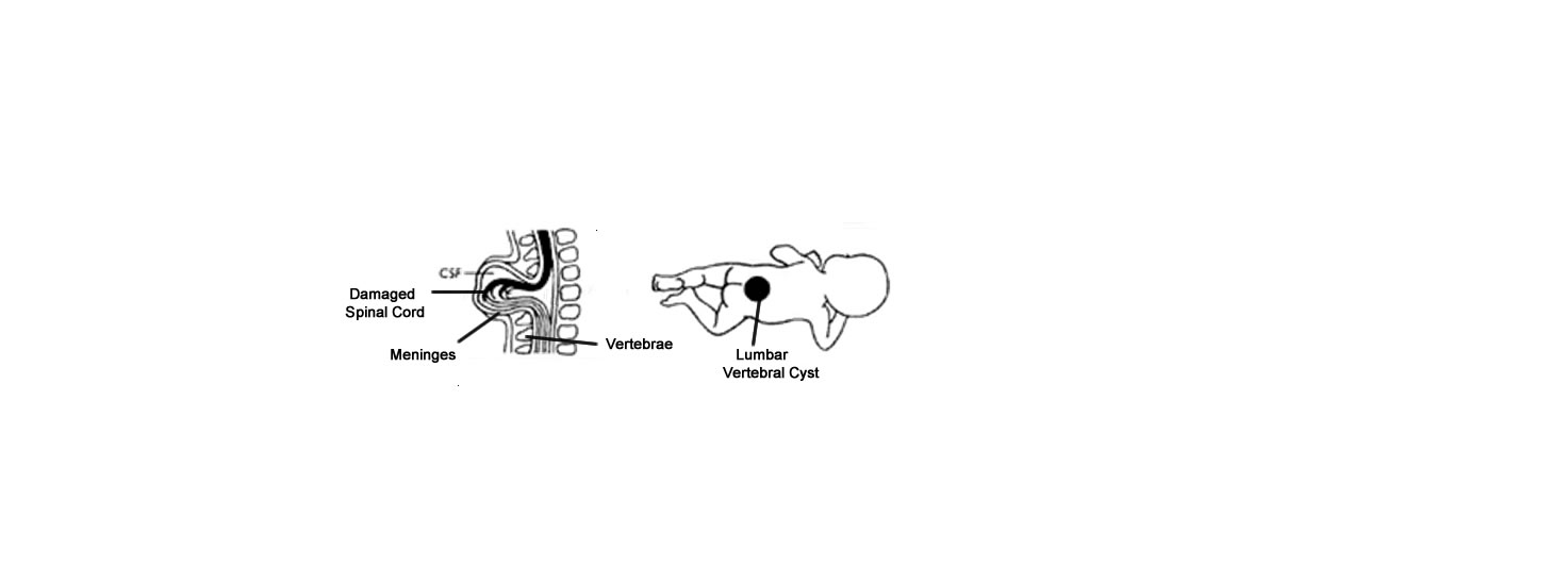 | 1,473 × 549 (24 KB) | Z3187802 (talk | contribs) | Modified image showing Spina Bifida Myelomeningocele characterized by split outer vertebrae with spinal cord and its meninges protruding from the divided vertebrae as a cyst. Commonly found at lumbar vertebral level |
You cannot overwrite this file.
File usage
The following file is a duplicate of this file (more details):
There are no pages that use this file.


