File:Migrating Neural Crest cells.png
From Embryology
Size of this preview: 579 × 600 pixels. Other resolution: 938 × 972 pixels.
Original file (938 × 972 pixels, file size: 53 KB, MIME type: image/png)
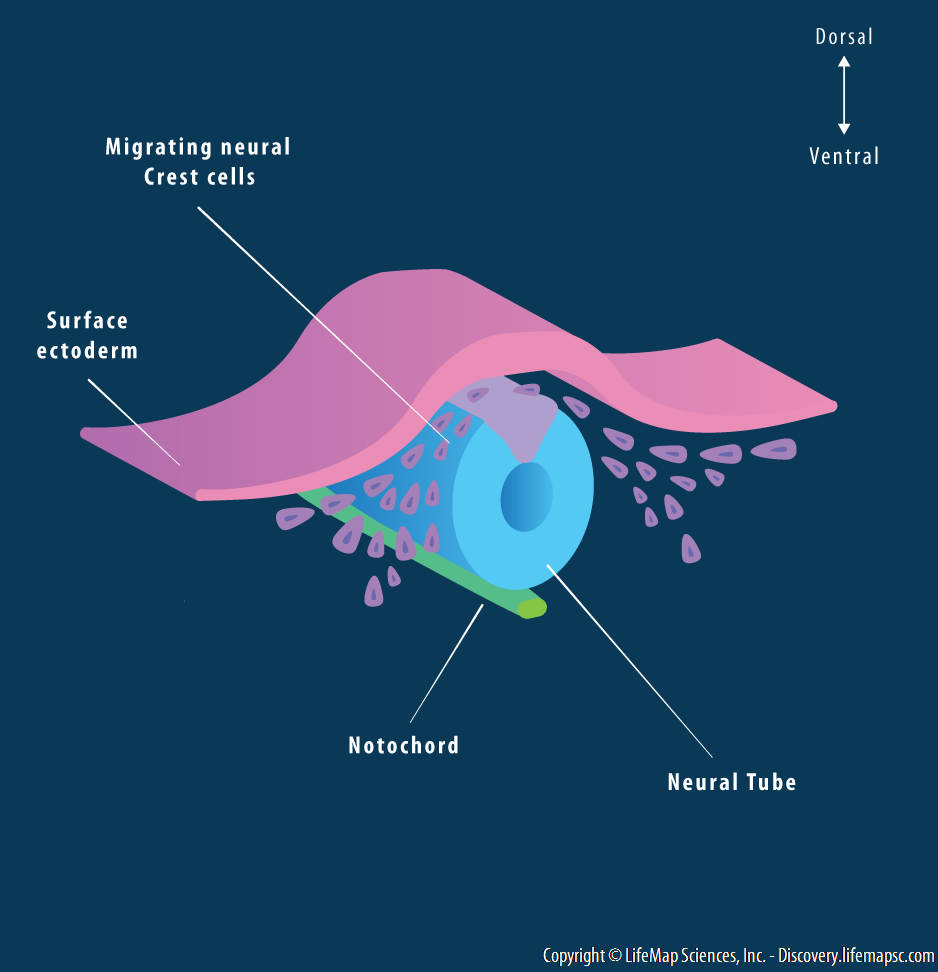
Both cranial (head) and trunk (body) neural crest populations are formed between the overlying surface ectoderm and the neural ectoderm during early vertebrate development. In response to distinct signals, these cells undergo epithelial-to-mesenchymal transition (EMT) and become migrating neural crest cells which contribute to different adult tissues.
File history
Yi efo/eka'e gwa ebo wo le nyangagi wuncin ye kamina wunga tinya nan
| Gwalagizhi | Nyangagi | Dimensions | User | Comment | |
|---|---|---|---|---|---|
| current | 11:41, 4 September 2018 | | 938 × 972 (53 KB) | Z5229281 (talk | contribs) | Both cranial (head) and trunk (body) neural crest populations are formed between the overlying surface ectoderm and the neural ectoderm during early vertebrate development. In response to distinct signals, these cells undergo epithelial-to-mesenchymal... |
You cannot overwrite this file.
File usage
The following page uses this file:


