2018 Group Project 3: Difference between revisions
(→Ears: added image Left and Right Cochlea.jpeg to page) |
(→Ears) |
||
| Line 16: | Line 16: | ||
Melanocytes are mainly present in the cochlea, vestibular organ and endolymphatic sac: | Melanocytes are mainly present in the cochlea, vestibular organ and endolymphatic sac: | ||
In the cochlea of humans: Melanocytes are found in the vascularised epithelial tissue in the intermediate layer of the stria vascularis and the modiolus, between the marginal and basal cell layers {{#pmid:3070525|PMID3070525}}. During development, marginal layer cuboidal epithelium develops processes which interdigitate with the intermediate melanocytes and basal cells {{#pmid: 2612372|PMID2612372}}. Based on studies performed on mice models, it is thought that melanocytes are important for the development of the endocochlear potential (EP), produced by strial cells. Intermediate melanocytes conduct K+, which plays an important role in sound conductance, as it flows from the endolymph into the ciliated epithelial cells of the ear via mechano-electrical channels. This influx of K+ is driven by a combination of the membrane potential of these ciliated epithelial cells, and the EP. A study on guinea pig models showed that blocking the K+ channels of melanocytes produces a lower EP {{#pmid: 8951443|PMID8951443}}. This study agrees with evidence showing that mice deficient in cochlea melanocytes have a lower EP, requiring a greater sound stimulus to produce an action potential {{#pmid: 7521050|PMID7521050}}. | In the cochlea of humans: Melanocytes are found in the vascularised epithelial tissue in the intermediate layer of the stria vascularis and the modiolus, between the marginal and basal cell layers {{#pmid:3070525|PMID3070525}}. During development, marginal layer cuboidal epithelium develops processes which interdigitate with the intermediate melanocytes and basal cells {{#pmid: 2612372|PMID2612372}}. Based on studies performed on mice models, it is thought that melanocytes are important for the development of the endocochlear potential (EP), produced by strial cells (Figure 1). Intermediate melanocytes conduct K+, which plays an important role in sound conductance, as it flows from the endolymph into the ciliated epithelial cells of the ear via mechano-electrical channels. This influx of K+ is driven by a combination of the membrane potential of these ciliated epithelial cells, and the EP. A study on guinea pig models showed that blocking the K+ channels of melanocytes produces a lower EP {{#pmid: 8951443|PMID8951443}}. This study agrees with evidence showing that mice deficient in cochlea melanocytes have a lower EP, requiring a greater sound stimulus to produce an action potential {{#pmid: 7521050|PMID7521050}}. | ||
[[File:Left and Right Cochlea.jpeg|400px]] | [[File:Left and Right Cochlea.jpeg|400px]] | ||
The cochlea of a Wv/Wv mutant showing the distribution of pigment within the Stria Vascularis {{#pmid:14909014|PMID14909014}} | Figure 1: The cochlea of a Wv/Wv mutant showing the distribution of pigment within the Stria Vascularis {{#pmid:14909014|PMID14909014}} | ||
===Eyes=== | ===Eyes=== | ||
Revision as of 09:10, 28 August 2018
| Projects 2018: 1 Adrenal Medulla | 3 Melanocytes | 4 Cardiac | 5 Dorsal Root Ganglion |
Project Pages are currently being updated (notice removed when completed)
Melanocytes
Introduction
History
Tissue Organ Structure and Function
Skin
Ears
Melanocytes are mainly present in the cochlea, vestibular organ and endolymphatic sac:
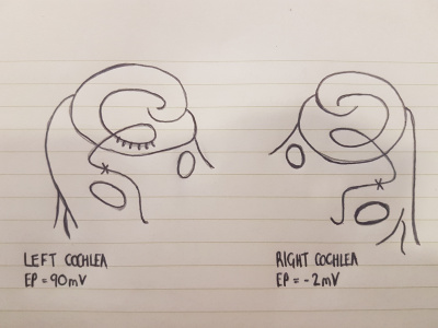
In the cochlea of humans: Melanocytes are found in the vascularised epithelial tissue in the intermediate layer of the stria vascularis and the modiolus, between the marginal and basal cell layers [1]. During development, marginal layer cuboidal epithelium develops processes which interdigitate with the intermediate melanocytes and basal cells [2]. Based on studies performed on mice models, it is thought that melanocytes are important for the development of the endocochlear potential (EP), produced by strial cells (Figure 1). Intermediate melanocytes conduct K+, which plays an important role in sound conductance, as it flows from the endolymph into the ciliated epithelial cells of the ear via mechano-electrical channels. This influx of K+ is driven by a combination of the membrane potential of these ciliated epithelial cells, and the EP. A study on guinea pig models showed that blocking the K+ channels of melanocytes produces a lower EP [3]. This study agrees with evidence showing that mice deficient in cochlea melanocytes have a lower EP, requiring a greater sound stimulus to produce an action potential [4].
Figure 1: The cochlea of a Wv/Wv mutant showing the distribution of pigment within the Stria Vascularis [5]
Eyes
Heart
CNS
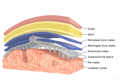
Melanocytes can also be found within the Central Nervous System, primarily in the leptomeninges [6], which consists of the inner two layers membranes of the meninges, the pia mater and arachnoid, which encapsulate the brain and the spinal cord. While its function within the meninges is unknown, they are essential to the health of the meninges, with their removal increases the risk of aseptic meningitis [7]. Furthermore, due to its receptivity to the same signalling molecules as neurons, scientists can study diseases afflicting the central nervous system using it as a model [8].
Leptomeninges comprises the arachnoid and pia mater
Embryonic Origins
Development Time Course
Molecular Mechanisms/ Factors/ Genes
Animal Models
Current Research
Abnormalities
Glossary
References
- ↑ Meyer zum Gottesberge AM. (1988). Physiology and pathophysiology of inner ear melanin. Pigment Cell Res. , 1, 238-49. PMID: 3070525
- ↑ Steel KP & Barkway C. (1989). Another role for melanocytes: their importance for normal stria vascularis development in the mammalian inner ear. Development , 107, 453-63. PMID: 2612372
- ↑ Takeuchi S, Kakigi A, Takeda T, Saito H & Irimajiri A. (1996). Intravascularly applied K(+)-channel blockers suppress differently the positive endocochlear potential maintained by vascular perfusion. Hear. Res. , 101, 181-5. PMID: 8951443
- ↑ Cable J, Huszar D, Jaenisch R & Steel KP. (1994). Effects of mutations at the W locus (c-kit) on inner ear pigmentation and function in the mouse. Pigment Cell Res. , 7, 17-32. PMID: 7521050
- ↑ DENIER A. (1951). [Microwaves of 12 cm; their biological and therapeutic effect]. J Radiol Electrol Arch Electr Medicale , 32, 664-5. PMID: 14909014
- ↑ Gudjohnsen SA, Atacho DA, Gesbert F, Raposo G, Hurbain I, Larue L, Steingrimsson E & Petersen PH. (2015). Meningeal Melanocytes in the Mouse: Distribution and Dependence on Mitf. Front Neuroanat , 9, 149. PMID: 26635543 DOI.
- ↑ Goldgeier MH, Klein LE, Klein-Angerer S, Moellmann G & Nordlund JJ. (1984). The distribution of melanocytes in the leptomeninges of the human brain. J. Invest. Dermatol. , 82, 235-8. PMID: 6699426
- ↑ Yaar M & Park HY. (2012). Melanocytes: a window into the nervous system. J. Invest. Dermatol. , 132, 835-45. PMID: 22158549 DOI.

