2016 Group Project 6: Difference between revisions
No edit summary |
No edit summary |
||
| Line 85: | Line 85: | ||
===Negative Regulation=== | ===Negative Regulation=== | ||
==Significance in Embryonic Development== | |||
===Cardiovascular Development=== | |||
TGF-B are cytokines involved in many biological processes. | |||
Genetic engineering and tissue explanation studies have revealed specific non-overlapping roles for TGF-B ligands and their signalling molecules in development (AND IN NORMAL FUNCTION OF CV SYSTEM IN ADULT – MENTION AT THE END. | |||
In the embryo, TGF-B appear to be involved in epithelial-mesenchymal transformations (EMT) during endocardial cushion formation, and in epicardial epithelial-mesenchymal transformations essential for coronary vasculature, ventricular myocardial development and compaction. | |||
In the adult, TGF-B are involved in cardiac hypertrophy, vascular remodelling and regulation of the renal renin-angiotensin system. | |||
It propagates the symmetrical embryonic cardiac tube into an asymmetrical four-chambered heart and requires considerable morphogenesis and remodelling processes. | |||
CV TGFB1-3 Expression – NEED THE PICTURE TO TALK ABOUT IT | |||
TGFB1 is expressed in the endocardium of the developing mouse. TGFB2-/- mice have obvious congenital cardiovascular defects, so its important to review its expression in the developing heart. In the blood vessels, TGFB1 is in the intima whereas TGFB2 and 3 are in the emdia and adventitia. | |||
TGFB2 message is found as early as embryonic day/E 7.25 (1) in the cardiogenic plate of the precardiac mesoderm and is later prominent in the myocardium of the aortic sac and outflow track regions. TGFB2 protein is found in the entire myocardium of the heart at the time when looping (morphogenesis when heart is formed by looping the tube into the shape of the heart). | |||
From E8.5-9.5 when the cushion formation (cells in development that play a role in the formation of the heart septa) process occurs, there is strong TGFB2 expression localised to the myocardium. (2ABDE) | |||
After cushion formation and EMT and before myocardialization of the endocardial cushion begins, there is strong TGFB2 expression in the OT myocardium and in the adjacent developing cushion mesenchyme. However as myocardialization occurs, the TGFB2 expression is reduced in the myocardium so that from E12.5 onwards, it is only expressed mainly in the mesenchyme of the cushion and OT septum. | |||
During myocardialization, TGFB2 expression remains high in the cushion mesenchyme of the OT septum (2GH). | |||
By E15.5, TGFB1 is now the most highly expressed isoform in the endocardial cells of the myocardium and TGFB2 is low, and the epicardium TGFB1 and 3 expression is higher than that of TGFB2 (2MNO). | |||
In general, TGFBs are expressed not in an overlapping fashion mainly. It is interesting to see that all three are expressed in the epicardium, and 2 but not 3 is expressed in the myocardium adjacent to the AV cushions. These observations are consistent with the fact that TGFB2-/- mice have abnormal pharyngeal arch artery remodelling and defective myocardialization of the OT septum. | |||
Specification of early cardiac precursor cells | |||
Cross talk between mesoderm and underlying endoderm is needed to form the early tubular heart. This cellular and molecular induction in the primary heart forming regions is important for the specification and differentiation of myocardial and endocardial precursor cells (2). Other endoderm-derived growth factors such as BMP2, FGF2 as well as TGFBS have been implicated in this process in the avian system (respiratory system delivering oxygen and removing carbon dioxide) (3). TGFB2 and TGFB receptors are expressed in the precardiac mesoderm along with BMP2 (4,5,6). Members of the TGG family can serve as inductive signals at the heart forming fields for the formation of myocardial and endocardial precursor cells. Members such as Activin, BMP, Nodal, Left and others have been found to be crucial for the establishment of embryonic asymmetry (7), and this asymmetry is in turn critical for heart development (8). | |||
1. https://www.ncbi.nlm.nih.gov/pubmed/7687212 | |||
2. https://www.ncbi.nlm.nih.gov/pubmed/10767078/ | |||
3. https://www.ncbi.nlm.nih.gov/pubmed/11322300/ | |||
4. https://www.ncbi.nlm.nih.gov/pubmed/7687212/ | |||
5. https://www.ncbi.nlm.nih.gov/pubmed/10767078/ | |||
6. https://www.ncbi.nlm.nih.gov/pubmed/10340759/ | |||
7. https://www.ncbi.nlm.nih.gov/pubmed/11836504/ | |||
8. https://www.ncbi.nlm.nih.gov/pubmed/11752633/ | |||
===Mammary Gland Development=== | |||
All three TGF-[beta] isoforms are expressed during all stages of mammary gland development except lactation (8). Mouse studies indicate key roles for TGF-[beta]s in establishing proper mammary gland architecture, regulating stem cell kinetics, maintaining the mammary epithelium in a functionally undifferentiated state, and inducing apoptosis in the involuting gland (2,9-12). The reader is referred to an earlier issue of this journal for additional comprehensive reviews (13). | |||
Importantly, TGF-[beta]s are potent inhibitors of the proliferation ofmammaryepithelial cells, both in vitro and in vivo, and the nature of the target cell may determine the type of TGF-[beta] response induced, as TGF-[beta] appears to inhibit proliferation in the ductal epithelial compartment, while inducing apoptosis without effects on proliferation in the alveolar compartment [reviewed (3)].This observation illustrates the general principle that the actions of TGF-[beta]s are very contextdependent, and are affected by cell type, environmental influences and cell history. | |||
TGF-[beta] Ligands. There are three closely-related mammalian isoforms of the TGF-[beta] ligand, and in vitro, TGF-[beta]1-3 generally elicit identical biological responses. Currently, it is thought that all three isoforms signal through the same T[beta]RII/T[beta]RI complex, though the possibility that there may be isoform selectivity in the nature and extent of post-receptor signaling has not yet been addressed. In the mammary gland, all three TGF-[beta] isoforms are expressed in the ductal epithelium at all stages of development except for lactation, but there is some isoform specificity in temporal and spatial expression patterns which may reflect isoform-specific roles [reviewed (2)]. For example, expression of epithelial TGF-[beta]2 is generally low but is upregulated during pregnancy, and TGF[beta]3 is the only isoform present in the endbud cap cells and myoepithelial cells. Uniquelyamongthe isoforms, TGF-[beta]1 is also present at high levels in the extracellular matrix that surrounds growth-quiescent ducts, consistent with a role for this isoform in the suppression of lateral budding once the ductal tree is established (2). However, it should be noted that TGF-[beta]s are synthesized as biologically latent forms, and that activation of the latent form is a critical regulatory step that must occur before the TGF-[beta]s can bind to their receptors (36). Most techniques for the localization of TGF-[beta] do not discriminate between active and latent TGF-[beta], and it is likely that the distribution of receptor-reactive TGF-[beta] is much less widespread than current techniques would imply. Indeed, using an elegant immunofluorescent technique, it has recently been shown that latent TGF-[beta] may be activated very locally on a cell-by-cell basis in the mammary gland, with activation in the nulliparous gland being confined primarily to a subpopulation of epithelial cells (4,37). Thus functional activation of the TGF-[beta] ligand/receptor signaling system should not be inferred from the mere co-localization of ligand and receptor, without additional information on ligand bioavailability. | |||
Activin and BMP Ligands. Activins and BMPs have been less extensively studied in the mammary gland. Activin [beta]B mRNA is expressed at all stages of mammary development, and a key role for stromallyderived activin [beta]Bin promoting ductal elongation and alveolar morphogenesis can be inferred from studies with the activin [beta]B knockout mouse (38). Activin [beta]AmRNAexpression was restricted to myoepithelial cells in studies of immunoaffinity-purified cell populations from the human breast (39). BMP-2 and BMP-4 mRNAs are expressed in both epithelial and stromal compartments during mammary gland development, withBMP-2expression being constitutive through development, while BMP-4 is down-regulated in late pregnancy and involution (4). | |||
Receptors and Signal Transduction Components. Systematic analysis of the expression of TGF-[beta] family receptor and signal transduction components in the mammary gland is still at an early stage. Table II summarizes what is currently known for the receptors. Immunohistochemical studies on the human breast showed that T[beta]RII is present in the ductal and lobular epithelial cells, but not the myoepithelial cells of the lobular units (40). In mouse, both T[beta]RII and T[beta]RI (Alk5) were expressed in both the mammary epithelium and the periductal stroma at all stages of development (9). These findings are consistent with transgenic mouse experiments suggesting that endogenous TGF-[beta] can act on both epithelium and stroma (9,11). In a study using human breast cells fractionated by immunoaffinity purification, myoepithelial cells, but not luminal epithelial cells or stromal cells, were shown to express mRNA for the activin/BMP receptor ActRII (39). This observation raises the possibility that TGF-[beta] may be more important in regulating stromal and luminal epithelial responses, with activin or BMPs regulating myoepithelial cells. Intriguingly, the ActRII gene was also expressed in all breast cancer cell lines studied and in microdissected invasive carcinoma cells, suggesting that inappropriate expression of ActRII in the mammary epithelium may play a role in tumorigenesis (39). Finally for BMPS, BMPRI-A (Alk3) mRNA was expressed most highly in blood vessels in the mouse mammary gland, with some expression in the periductal stroma at all stages of development, and also in the epithelium of involuting alveoli (9). The expression in blood vessels is consistent with known roles for the BMP pathway in the developing embryonic vasculature (21). In contrast, ActRI (Alk2) was predominantly localized to the mammary epithelium (9). Since ActRI is now thought to be on the BMP response pathway (41), this finding suggests there will be specific effects ofBMPson the mammary epithelium. | |||
So far there are no published reports on Smad expression in the normal mammary gland. However, we find mRNAs for Smads1-5 in the mouse mammary gland at all stages of development [Y. Yang and L. Wakefield, in preparation], which would support the concept that both TGF-[beta]/activin and BMP signal transduction pathways are operational in the mammary gland. Details of the distribution of the Smads between different cellular compartments in the mammary gland remain to be established. In a small study of six human breast cancer cell lines, Smads2 and 3 were found in all lines, and Smad4 in all but one (42), which suggests these three Smads are probably present in the normal epithelium. | |||
===Development of hair follicles/teeth/submandibular gland=== | |||
===Vascular biology and dysfunction=== | |||
===Maintenance of pluripotency in hESC=== | |||
===Role in cancer=== | |||
===Formation of digits from limb digits=== | |||
===Formation of the palate=== | |||
==Current Research== | ==Current Research== | ||
Revision as of 01:44, 20 October 2016
| 2016 Student Projects | ||||
|---|---|---|---|---|
| Signalling: 1 Wnt | 2 Notch | 3 FGF Receptor | 4 Hedgehog | 5 T-box | 6 TGF-Beta | ||||
| 2016 Group Project Topic - Signaling in Development
OK you are now in a group, add a topic with your student signature to the group page. | ||||
| This page is an undergraduate science embryology student project and may contain inaccuracies in either descriptions or acknowledgements. | ||||
| Group Assessment Criteria |
|---|
 Science Student Projects Science Student Projects
|
| More Information on Assessment Criteria | Science Student Projects |
Transforming Growth Factor (TGF) Beta Signalling Pathway
Introduction
Transforming Growth Factor (TGF) beta is a multifunctional peptide/cytokine that controls proliferation, cellular differentiation, angiogenesis and other functions in various cell types. TGF-beta plays a dominant part in the development of the embryo and adult organism, as well as cell growth, immune function and hormone secretion.
TGF-beta belongs to the Transforming Growth Factor superfamily, a large group of structurally connected cell regulatory proteins. It consists of TGF-beta 1, 2 and 3, Activins, Inhibins, Lefty, Nodal, Growth Differentiation Factors (GDFs), Bone Morphogenetic Proteins (BMPs), Glial-derived Neurotrophic Factors (GDNFs) and Mullierian Inhibiting Substance (MIS). This site will focus on the TGF-beta family. TGF betas are involved in embryogenesis. During development of the embryo, members of the TGF-beta family are essential for bone and cartilage formation, mesoderm induction and patterning and dorso-ventral patterning.
Canonical pathway
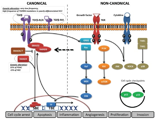
In the canonical pathway, the three TGF-β ligand isoforms, TGF-β1, TGF-β2, and TGF-β3, are synthesized as precursors and stick together to form the dormant TGF-β complex. It is then secreted, then following extracellular activation, TGF-β ligands bind to the membranous TGF-β type III receptor or the TGF-β type II receptor (TGF-βRII) homodimers with high affinity. TGF-βRII binding allows dimerization with TGF-β type I receptor (TGF-βRI) homodimers, activation of the TGF-βRI kinase domain and signal transduction via phosphorylation of the C-terminus of receptor-regulated SMADs (R-SMAD), SMAD2 and SMAD3. The TGF-βR dimer then forms a heterotrimeric complex with SMAD4 which translocates and accumulates in the nucleus. TGF-β dependent signalling can activate or repress hundreds of target genes through the interaction of SMADs with various transcription factors (TF). SMAD activities are regulated through several mechanisms: SMAD2/3 nucleocytoplasmic shuttling, binding to anchor proteins such as SARA, phosphorylation (e.g., by ERK, JNK, and p38 MAPK), Smurf (SMAD-ubiquitination-regulatory factor)-dependent degradation, or via expression of inhibitory SMAD6 and SMAD7.
Non-Canonical pathway
In the non-canonical pathway, TGF-β signalling activates SMAD-independent pathways such as PI3K/AKT, MAPK pathways (ERK, JNK, and p38 MAPK) as well as NF-κB, Rho/Rac1, Cdc42, FAK, Src, Abl[142]. Moreover, transversal signalling, especially at the SMAD level, allows TGF-β pathway activation to integrate signals from integrins, Notch, Wnt, TNF-α, or EGF-dependent pathways as well as signals from cellular processes such as the cell cycle or apoptosis machineries. The TGF-β signalling pathway thus has pleiotropic functions regulating cell growth, differentiation, apoptosis, cell motility, extracellular matrix production, angiogenesis and cellular immune response. reworddddddddd
Process of TGF-beta signalling pathway
<html5media width="560" height="315">https://www.youtube.com/watch?v=GuKjUearIUI</html5media>
The Transforming Growth Factor (TGF) beta signalling pathway is required for regulation of a large number of cellular processes such as cell proliferation, invasion and inflammation. It is also activated mitogen activated protein kinase signalling. There are two main routes in TGF-Beta signalling; the SMAD dependant pathway and SMAD independent pathway.
SMAD dependant TGF-beta signalling
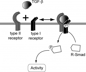
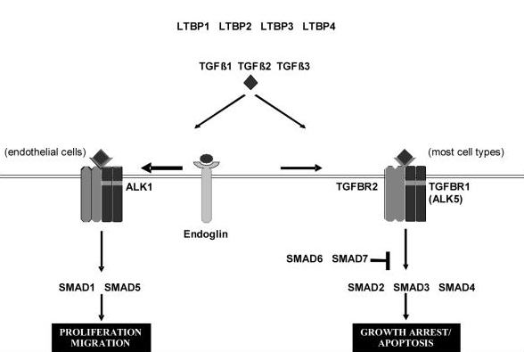
TGF beta superfamily ligands form dimers that bind to heterodimeric receptor complexes composed of two type I and two type II transmembrane receptor subunits with serine/threonine kinase domains. Following ligand binding, the Type II receptor (TGF-beta RII) phosphorylates and activates the Type I receptor (TGF-beta RI). In most cell types, this leads to recruitment and phosphorylation of SMAD2 and SMAD3. (SMAD is a family of gene regulatory proteins). SMAD1 and SMAD5 can be activated by TGF-beta signalling in some cell types depending on the Type I receptor that is expressed. Activated SMAD proteins associate with SMAD4 and translocate to the nucleus, where they accumulate (and act as transcription factors and participate in the regulation of target gene expression). They recruit additional transcriptional regulators, including DNA-binding transcription factors, co-activators, co-repressors and chromatin remodeling factors, that control the expression of numerous target genes.This initiates a SMAD-dependent signalling cascade that induces or represses transcriptional activity. SMADs are widely expressed in most adult tissue and cell types indicating that the TGF-beta signalling pathway is ubiquitous. Differential expression of these factors may be responsible for some of the cell type-specific responses to TGF-beta.
Note: Type I cytokine receptors are also transmembrane receptors expressed on the surface of cells. They recognize and respond to cytokines with four alpha helical strands. Type II cytokine receptors are transmembrane proteins that are expressed on the surface of certain cells. The difference between Type I and Type II receptors is that Type II receptors do not possess the signature sequence WSXWS, which is a characteristic of Type I receptors.
(Tabulate how it is involved in the various processes of embryology)
SMAD independant TGF-beta signalling
Rather than SMAD-mediated transciption TGF-β also has the potential to activate other signalling cascades for example the Erk, JNK and p38 MAPK kinase pathways. In some cases these pathways exhibit activation with slow kinetics which indicates SMAD-dependant mechanics, however there has also been rapid activation cases (5-15mins) suggesting independence from transcription mechanisms. Studies carried out with SMAD4 deficient cells and dominant-negative SMADS provide evidence that the MAPK pathway activation is independent from SMADS, as well as this it has be found that p38 MAPK signalling was activated in response to mutated TGF- β type 1 receptors, which were defective in SMAD activation[2].
The precise mechanisms and biological consequences of these SMAD-Independent pathways (Erk, JNK, p38 MAPK) are currently poorly characterized. Ras is implicated in TGF- β induced Erk signalling as there is rapid activation of Ras by TGF- β in epithelial cells. The JNK and p38 MAPK signalling are activated by various MAPK kinase kinases (MAPKKK) TGF- β kinase 1 (TAK 1) receptor is a MAPKKK family member. Further research and identification of various interactions between the small signalling molecules and receptor proteins will provide additional insight into the precise mechanism behind the activation of MAPK pathways by TGF- β ligands [2].
Regulation of the pathway and factors affecting it
Signalling mechanisms by TGF-β like factors are regulated in both negative and positive fashions, these are all tightly controlled through a multitude of mechanisms at extracellular, membrane, cytoplasmic and all the way to nuclear levels. Positive regulation is required to amplify signalling from TGF-β like factors, while negative regulation is important for the termination and restriction of signalling usually occurring through the mechanism of a feedback loop. There is also additional regulation of TGF-β like factors via cross-talk with other signal transduction pathways such as MAPK and JAK/STAT pathways[3].
Positive Regulation
The positive regulation of TGF-β specifically the induction of ligands and their signalling components often is triggered by the action TGF-β-like factors themselves. For example NODAL, a secretory protein of the TGF-β superfamily which plays a role in early embryogenesis and acts through activin receptors and SMAD2 is induced by nodal signalling itself[3].
Negative Regulation
Significance in Embryonic Development
Cardiovascular Development
TGF-B are cytokines involved in many biological processes. Genetic engineering and tissue explanation studies have revealed specific non-overlapping roles for TGF-B ligands and their signalling molecules in development (AND IN NORMAL FUNCTION OF CV SYSTEM IN ADULT – MENTION AT THE END. In the embryo, TGF-B appear to be involved in epithelial-mesenchymal transformations (EMT) during endocardial cushion formation, and in epicardial epithelial-mesenchymal transformations essential for coronary vasculature, ventricular myocardial development and compaction. In the adult, TGF-B are involved in cardiac hypertrophy, vascular remodelling and regulation of the renal renin-angiotensin system.
It propagates the symmetrical embryonic cardiac tube into an asymmetrical four-chambered heart and requires considerable morphogenesis and remodelling processes.
CV TGFB1-3 Expression – NEED THE PICTURE TO TALK ABOUT IT TGFB1 is expressed in the endocardium of the developing mouse. TGFB2-/- mice have obvious congenital cardiovascular defects, so its important to review its expression in the developing heart. In the blood vessels, TGFB1 is in the intima whereas TGFB2 and 3 are in the emdia and adventitia. TGFB2 message is found as early as embryonic day/E 7.25 (1) in the cardiogenic plate of the precardiac mesoderm and is later prominent in the myocardium of the aortic sac and outflow track regions. TGFB2 protein is found in the entire myocardium of the heart at the time when looping (morphogenesis when heart is formed by looping the tube into the shape of the heart). From E8.5-9.5 when the cushion formation (cells in development that play a role in the formation of the heart septa) process occurs, there is strong TGFB2 expression localised to the myocardium. (2ABDE) After cushion formation and EMT and before myocardialization of the endocardial cushion begins, there is strong TGFB2 expression in the OT myocardium and in the adjacent developing cushion mesenchyme. However as myocardialization occurs, the TGFB2 expression is reduced in the myocardium so that from E12.5 onwards, it is only expressed mainly in the mesenchyme of the cushion and OT septum. During myocardialization, TGFB2 expression remains high in the cushion mesenchyme of the OT septum (2GH). By E15.5, TGFB1 is now the most highly expressed isoform in the endocardial cells of the myocardium and TGFB2 is low, and the epicardium TGFB1 and 3 expression is higher than that of TGFB2 (2MNO). In general, TGFBs are expressed not in an overlapping fashion mainly. It is interesting to see that all three are expressed in the epicardium, and 2 but not 3 is expressed in the myocardium adjacent to the AV cushions. These observations are consistent with the fact that TGFB2-/- mice have abnormal pharyngeal arch artery remodelling and defective myocardialization of the OT septum.
Specification of early cardiac precursor cells Cross talk between mesoderm and underlying endoderm is needed to form the early tubular heart. This cellular and molecular induction in the primary heart forming regions is important for the specification and differentiation of myocardial and endocardial precursor cells (2). Other endoderm-derived growth factors such as BMP2, FGF2 as well as TGFBS have been implicated in this process in the avian system (respiratory system delivering oxygen and removing carbon dioxide) (3). TGFB2 and TGFB receptors are expressed in the precardiac mesoderm along with BMP2 (4,5,6). Members of the TGG family can serve as inductive signals at the heart forming fields for the formation of myocardial and endocardial precursor cells. Members such as Activin, BMP, Nodal, Left and others have been found to be crucial for the establishment of embryonic asymmetry (7), and this asymmetry is in turn critical for heart development (8).
1. https://www.ncbi.nlm.nih.gov/pubmed/7687212
2. https://www.ncbi.nlm.nih.gov/pubmed/10767078/
3. https://www.ncbi.nlm.nih.gov/pubmed/11322300/
4. https://www.ncbi.nlm.nih.gov/pubmed/7687212/
5. https://www.ncbi.nlm.nih.gov/pubmed/10767078/
6. https://www.ncbi.nlm.nih.gov/pubmed/10340759/
7. https://www.ncbi.nlm.nih.gov/pubmed/11836504/
8. https://www.ncbi.nlm.nih.gov/pubmed/11752633/
Mammary Gland Development
All three TGF-[beta] isoforms are expressed during all stages of mammary gland development except lactation (8). Mouse studies indicate key roles for TGF-[beta]s in establishing proper mammary gland architecture, regulating stem cell kinetics, maintaining the mammary epithelium in a functionally undifferentiated state, and inducing apoptosis in the involuting gland (2,9-12). The reader is referred to an earlier issue of this journal for additional comprehensive reviews (13). Importantly, TGF-[beta]s are potent inhibitors of the proliferation ofmammaryepithelial cells, both in vitro and in vivo, and the nature of the target cell may determine the type of TGF-[beta] response induced, as TGF-[beta] appears to inhibit proliferation in the ductal epithelial compartment, while inducing apoptosis without effects on proliferation in the alveolar compartment [reviewed (3)].This observation illustrates the general principle that the actions of TGF-[beta]s are very contextdependent, and are affected by cell type, environmental influences and cell history. TGF-[beta] Ligands. There are three closely-related mammalian isoforms of the TGF-[beta] ligand, and in vitro, TGF-[beta]1-3 generally elicit identical biological responses. Currently, it is thought that all three isoforms signal through the same T[beta]RII/T[beta]RI complex, though the possibility that there may be isoform selectivity in the nature and extent of post-receptor signaling has not yet been addressed. In the mammary gland, all three TGF-[beta] isoforms are expressed in the ductal epithelium at all stages of development except for lactation, but there is some isoform specificity in temporal and spatial expression patterns which may reflect isoform-specific roles [reviewed (2)]. For example, expression of epithelial TGF-[beta]2 is generally low but is upregulated during pregnancy, and TGF[beta]3 is the only isoform present in the endbud cap cells and myoepithelial cells. Uniquelyamongthe isoforms, TGF-[beta]1 is also present at high levels in the extracellular matrix that surrounds growth-quiescent ducts, consistent with a role for this isoform in the suppression of lateral budding once the ductal tree is established (2). However, it should be noted that TGF-[beta]s are synthesized as biologically latent forms, and that activation of the latent form is a critical regulatory step that must occur before the TGF-[beta]s can bind to their receptors (36). Most techniques for the localization of TGF-[beta] do not discriminate between active and latent TGF-[beta], and it is likely that the distribution of receptor-reactive TGF-[beta] is much less widespread than current techniques would imply. Indeed, using an elegant immunofluorescent technique, it has recently been shown that latent TGF-[beta] may be activated very locally on a cell-by-cell basis in the mammary gland, with activation in the nulliparous gland being confined primarily to a subpopulation of epithelial cells (4,37). Thus functional activation of the TGF-[beta] ligand/receptor signaling system should not be inferred from the mere co-localization of ligand and receptor, without additional information on ligand bioavailability.
Activin and BMP Ligands. Activins and BMPs have been less extensively studied in the mammary gland. Activin [beta]B mRNA is expressed at all stages of mammary development, and a key role for stromallyderived activin [beta]Bin promoting ductal elongation and alveolar morphogenesis can be inferred from studies with the activin [beta]B knockout mouse (38). Activin [beta]AmRNAexpression was restricted to myoepithelial cells in studies of immunoaffinity-purified cell populations from the human breast (39). BMP-2 and BMP-4 mRNAs are expressed in both epithelial and stromal compartments during mammary gland development, withBMP-2expression being constitutive through development, while BMP-4 is down-regulated in late pregnancy and involution (4). Receptors and Signal Transduction Components. Systematic analysis of the expression of TGF-[beta] family receptor and signal transduction components in the mammary gland is still at an early stage. Table II summarizes what is currently known for the receptors. Immunohistochemical studies on the human breast showed that T[beta]RII is present in the ductal and lobular epithelial cells, but not the myoepithelial cells of the lobular units (40). In mouse, both T[beta]RII and T[beta]RI (Alk5) were expressed in both the mammary epithelium and the periductal stroma at all stages of development (9). These findings are consistent with transgenic mouse experiments suggesting that endogenous TGF-[beta] can act on both epithelium and stroma (9,11). In a study using human breast cells fractionated by immunoaffinity purification, myoepithelial cells, but not luminal epithelial cells or stromal cells, were shown to express mRNA for the activin/BMP receptor ActRII (39). This observation raises the possibility that TGF-[beta] may be more important in regulating stromal and luminal epithelial responses, with activin or BMPs regulating myoepithelial cells. Intriguingly, the ActRII gene was also expressed in all breast cancer cell lines studied and in microdissected invasive carcinoma cells, suggesting that inappropriate expression of ActRII in the mammary epithelium may play a role in tumorigenesis (39). Finally for BMPS, BMPRI-A (Alk3) mRNA was expressed most highly in blood vessels in the mouse mammary gland, with some expression in the periductal stroma at all stages of development, and also in the epithelium of involuting alveoli (9). The expression in blood vessels is consistent with known roles for the BMP pathway in the developing embryonic vasculature (21). In contrast, ActRI (Alk2) was predominantly localized to the mammary epithelium (9). Since ActRI is now thought to be on the BMP response pathway (41), this finding suggests there will be specific effects ofBMPson the mammary epithelium.
So far there are no published reports on Smad expression in the normal mammary gland. However, we find mRNAs for Smads1-5 in the mouse mammary gland at all stages of development [Y. Yang and L. Wakefield, in preparation], which would support the concept that both TGF-[beta]/activin and BMP signal transduction pathways are operational in the mammary gland. Details of the distribution of the Smads between different cellular compartments in the mammary gland remain to be established. In a small study of six human breast cancer cell lines, Smads2 and 3 were found in all lines, and Smad4 in all but one (42), which suggests these three Smads are probably present in the normal epithelium.
Development of hair follicles/teeth/submandibular gland
Vascular biology and dysfunction
Maintenance of pluripotency in hESC
Role in cancer
Formation of digits from limb digits
Formation of the palate
Current Research
Abnormalities of the TGF-Beta Pathway
Mutations or deletion of the TGF-beta 1 or TGF-beta RII gene have been associated with multiple syndromes. In mice, defects have been found in haematopoiesis, vasculogenesis and endothelial differentiation of extra embryonic tissues, while knockout mice for SMAD2 or SMAD4 genes are more likely to have spontaneous tumour development and excessive inflammatory responses. In humans, various diseases have been linked to the mutation of the TGF-beta RII gene and SMAD4 mutation is genetically responsible for familial juvenile polyposis, an autosomal dominant disease characterized by predisposition to gastrointestinal polyps and cancers.
Alterations of this signalling pathway are common in cancer. Accessory proteins such as soluble or membrane-bound regulators or co-receptors can also affect TGF-beta signalling.
Further Reading
Glossary
Apoptosis - cell death which occurs as a normal and controlled part of an organism's growth or development
Cytokine - a broad and loose category of small proteins that are important in cell signalling
Ligands - a molecule that binds to a larger molecule
CCL-64 - mink lung epithelial cell
History
From the early stages of TGF beta to the present day, research and studies on the signaling pathway have radically increased. SMAD signaling and the three receptors for TGF-beta are two of the many fields of interest regarding the topic. In medicine and specific areas such as cancer, cardiovascular disease and inflammatory bowel disease, there are numerous alternatives for drugs that can either heighten or suppress the activity of TGF-beta.
| 1970s | It was believed that the growth of normal cells was mainly controlled by the interaction between various polypeptide hormones and hormone-like growth factors that were found in tissue fluids. Numerous new polypeptide growth factors had just been classified in cellular extracts, blood and serum.
It was also noted that malignant cells were not prone to be affected by all the same growth controls compared to normal cells and needed a lesser amount of these exogenous growth factors for optimal growth and multiplication. |
| Early 1980s | Anita Roberts found that SGF was not a particular substance, but a combination of two or more elements. One fraction was named “transforming growth factor-a”, the embryonic form of the epidermal growth factor (EGF) found in the salivary gland of an adult. The other fraction showed no rivalry with EGF in a receptor binding assay, but had the striking quality of generating the growth of various large colonies of NRK cells and was called “transforming growth factor-b”.
The theory that TGF’s were cancer-specific was proved inaccurate. In vivo studies established the initial hypothesis that one of the functions of TGF-beta in normal tissues was to be involved in the process of wound healing. |
| 1980 | The “autocrine secretion” hypothesis was developed. It proposed that the supposed transformed cell should produce the transforming polypeptide and have its own functional cellular receptors. This model implied that the endogenous production of growth-promoting polypeptides by a transformed cell would minimize its own need for an exogenous supply of alike growth factors. |
| 1984 | Moses and colleagues made a significant finding that TGF-beta could hinder cell growth if an suitable reader cell such as CCL-64 was used
The first receptor binding assay was published |
| 1985 | TGF-beta1 was cloned by Derynck and colleagues at Genentech
It was shown that TGF-beta could be multifunctional in the exact cells in which it was assayed, based on the context of the assay |


